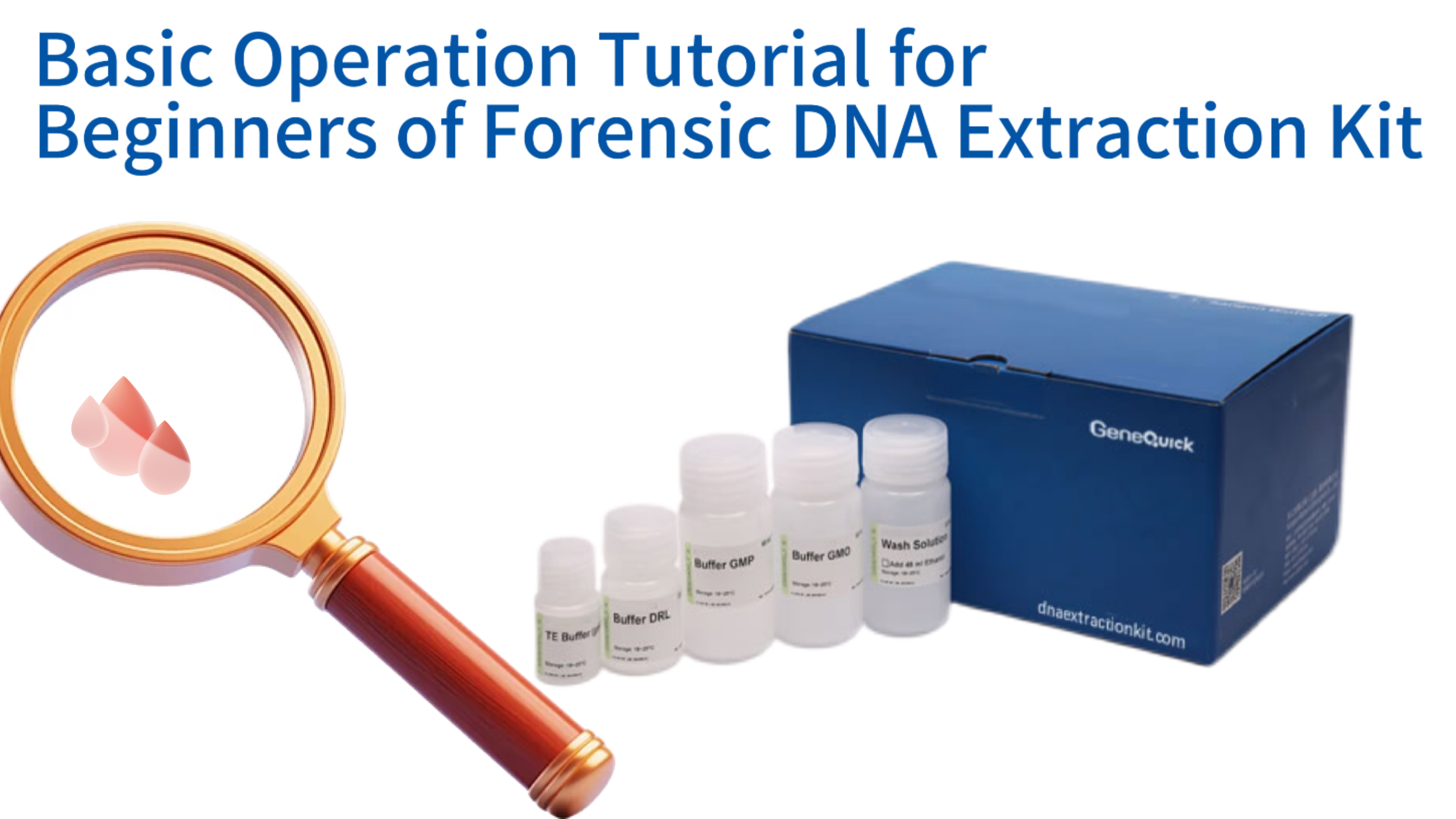
Basic Operation Tutorial for Beginners of Forensic DNA Extraction Kit
Entering the field of forensic DNA analysis requires a solid understanding of the fundamental techniques used to obtain genetic material from crime scene samples. A forensic DNA extraction kit is specifically designed to address the unique challenges posed by trace evidence, degraded samples, and the presence of PCR inhibitors. This tutorial provides a comprehensive, step‑by‑step guide for beginners, explaining the scientific principles behind each phase of the extraction process and offering practical advice for handling various forensic specimens. By mastering these procedures, users can ensure the recovery of high‑quality DNA suitable for downstream applications such as STR profiling and sequencing, while maintaining the chain of custody and preventing contamination. The following sections break down the entire workflow, from initial workspace preparation to final quality control, empowering new analysts to perform reliable extractions with confidence.
Understanding Forensic DNA Extraction: Principles and Challenges
Impact of Inhibitor Concentration on PCR Amplification Efficiency
Comparison of Forensic DNA Extraction Technologies
| Feature | Spin-Column Chromatography | Magnetic-Bead Separation |
|---|---|---|
| Equipment Requirement | Standard Centrifuge | Magnetic Separator |
| Automation Potential | Low | High |
| Hands-on Time | Moderate | Low |
| Suitability for Beginners | High | Moderate |
| Cost per Sample | Low | Moderate |
The Unique Nature of Forensic Samples
Forensic samples are rarely pristine. They can be exposed to environmental insults like humidity, UV light, or microbial degradation, all of which can damage DNA molecules. Moreover, substrates such as denim, wood, or soil introduce compounds that co‑extract with DNA and inhibit polymerase chain reaction (PCR). For instance, humic acids from soil are notorious for interfering with Taq polymerase activity. Therefore, a forensic extraction kit must not only lyse cells efficiently but also remove these inhibitors to produce a pure DNA preparation. Studies published in the International Journal of Legal Medicine have shown that even a 1% residual inhibitor concentration can reduce PCR amplification efficiency by over 50%, underscoring the need for rigorous purification.
Another challenge is the low template number often encountered in forensic work. Touch DNA, derived from shed skin cells, may contain less than 100 picograms of total DNA. In such cases, every step of the extraction must be optimized to minimise losses. Silica‑based methods, whether in spin‑column or magnetic‑bead format, are favoured because they bind DNA reversibly, allowing concentrated elution in small volumes. This design maximises the final DNA concentration, which is crucial for obtaining complete short tandem repeat (STR) profiles from low‑level samples.
Core Technologies in Forensic Kits
Two primary technologies dominate the forensic extraction landscape: spin‑column chromatography and magnetic‑bead separation. Spin‑column kits utilise a silica membrane housed in a microcentrifuge tube; under high salt conditions, DNA binds to the silica while contaminants pass through. After washing, low‑salt buffer releases the purified DNA. This method is straightforward and requires only a standard centrifuge, making it accessible to most laboratories. Magnetic‑bead technology, on the other hand, employs paramagnetic particles coated with silica or carboxyl groups. DNA binds to the beads in the presence of a chaotropic salt, and a magnetic rack separates the bead‑DNA complex from the liquid phase. Bead‑based protocols are easily automated and can be scaled for high‑throughput processing, which is increasingly important in busy forensic facilities.
Both technologies have been validated extensively. A comparative study in Forensic Science International: Genetics evaluated several commercial kits and found that silica‑based methods consistently yielded DNA suitable for multiplex STR amplification. The choice between column and beads often depends on laboratory workflow, sample throughput, and personal preference. For beginners, spin‑column kits may offer a more intuitive hands‑on experience, while magnetic‑bead systems provide flexibility for automated platforms. Regardless of the format, the underlying chemistry relies on the same principle: selective adsorption of nucleic acids onto a solid phase in the presence of chaotropic agents such as guanidinium thiocyanate.
Key Reagents and Their Functions
A forensic DNA extraction kit typically includes several specialised buffers. The lysis buffer contains a detergent (e.g., SDS or Triton X‑100) to disrupt cell membranes and a proteinase K solution to digest histones and other DNA‑binding proteins. Proteinase K is particularly important for forensic samples because it also inactivates nucleases that might otherwise degrade the DNA. The binding buffer is a high‑salt, chaotropic solution that facilitates DNA adsorption to the silica matrix. It often contains guanidine hydrochloride or guanidinium thiocyanate, which denature proteins and help release DNA from cellular debris. Wash buffers usually contain ethanol or isopropanol to remove residual contaminants while keeping the DNA bound to the solid phase. Finally, the elution buffer is a low‑ionic‑strength Tris‑EDTA solution (typically pH 8.0–8.5) that releases the purified DNA from the silica. Some kits also include an optional carrier RNA or poly‑A carrier to improve recovery from low‑input samples, a feature especially valuable for forensic applications involving trace DNA.
Each reagent’s composition is carefully balanced to maximise yield and purity. For example, the pH of the elution buffer is critical because DNA elutes more efficiently at slightly alkaline conditions. If the pH drops below 7.0, the DNA may remain bound to the silica, resulting in poor recovery. Manufacturers perform extensive quality control to ensure batch‑to‑batch consistency, which is essential for forensic laboratories that must adhere to accreditation standards such as ISO 17025.
Importance of Purity and Integrity for Downstream Analysis
The ultimate success of any forensic investigation hinges on the quality of the extracted DNA. Purity is commonly assessed by spectrophotometric ratios: A260/A280 should be between 1.8 and 2.0, indicating minimal protein or phenol contamination, while A260/A230 should exceed 2.0, reflecting low levels of chaotropic salts or carbohydrates. Deviations from these values can lead to failed amplifications or artefactual peaks in STR electropherograms. DNA integrity, or the average fragment size, is equally important. Degraded DNA produces partial profiles and allele drop‑out, compromising the evidential value. Forensic extraction kits are designed to preserve long fragments by using gentle lysis conditions and avoiding vigorous vortexing, which can shear high‑molecular‑weight DNA. A 2020 review in Wiley Interdisciplinary Reviews: Forensic Science highlighted that kits optimised for forensic use consistently outperform generic molecular biology kits in recovering amplifiable DNA from challenging substrates like bones and teeth.
Preparing for the Extraction: Workspace and Sample Handling
Essential Equipment for Forensic DNA Extraction
| Equipment Type | Minimum Specification |
|---|---|
| Microcentrifuge | 14,000 × g capacity |
| Pipettes | 2 µL to 1000 µL range |
| Heating Block/Water Bath | 56°C and 70°C settings |
| Magnetic Separator | For bead-based protocols |
| Freezer | -20°C (minimum) |
Setting Up a Clean Work Environment
Forensic DNA laboratories operate under strict protocols to prevent cross‑contamination between samples. The extraction area should be physically separated from post‑PCR areas, and unidirectional workflow must be enforced. Before beginning, the analyst should disinfect all work surfaces with a 10% bleach solution followed by 70% ethanol to remove any residual DNA and nucleases. Dedicated equipment, including pipettes, tube racks, and centrifuges, should be reserved for pre‑amplification steps. Many laboratories also use UV‑irradiation cabinets or HEPA‑filtered hoods to create a DNA‑free zone for handling low‑template samples. Personal protective equipment, such as lab coats, gloves, and face masks, must be worn at all times, and gloves should be changed frequently, especially after touching potentially contaminated surfaces.
It is also wise to include a reagent blank (extraction control) alongside each batch of samples. This control contains all the kit reagents but no sample material; if DNA is detected in the blank, it signals contamination during the extraction process. Such controls are mandated by forensic quality standards like ISO 18385, which specifically addresses the requirements for products used in forensic DNA analysis. Maintaining a clean environment and including appropriate controls are the first lines of defence against erroneous results that could mislead an investigation.
Essential Equipment and Consumables
While the extraction kit itself provides the chemical reagents, the analyst must supply various pieces of equipment. A microcentrifuge capable of at least 14,000 × g is required for spin‑column protocols, and a magnetic separator is necessary for bead‑based methods. Adjustable pipettes covering volumes from 2 µL to 1000 µL, together with aerosol‑resistant filter tips, are indispensable for accurate liquid handling. Heating blocks or water baths set to 56°C and 70°C are used for lysis and elution steps. Vortex mixers help resuspend pellets, but care must be taken to avoid excessive shearing of DNA. For sample storage, –20°C freezers are standard, while some laboratories also use –80°C for long‑term preservation. All consumables, including microcentrifuge tubes and pipette tips, should be certified DNase‑free and RNase‑free. Many forensic labs additionally pre‑sterilise plasticware by autoclaving to eliminate any contaminating nucleases.
Beyond the basics, specialised items may be needed for certain sample types. For instance, processing bone or tooth samples often requires a freezer mill or a drill to produce fine powder, while swabs may be air‑dried before extraction. Having these tools ready before starting the protocol prevents unnecessary delays and reduces the chance of sample degradation during handling.
Sample Receipt and Documentation
Forensic evidence arrives with a chain‑of‑custody form that must be meticulously maintained. Upon receipt, the analyst checks the packaging for integrity, records the condition, and assigns a unique laboratory identifier. Photographs may be taken to document the state of the evidence. For liquid samples like blood, the volume is noted; for stains, the approximate size and substrate are recorded. This documentation is critical because any discrepancies could later be challenged in court. After documentation, a portion of the sample is subsampled for extraction. The remainder is returned to secure storage. It is advisable to retain a portion of the original sample whenever possible, as it may be needed for re‑analysis or confirmation by an independent laboratory.
When handling trace evidence, extreme care is taken to avoid loss. For example, when cutting a stained fabric, sterile scalpels or scissors are used, and the cutting surface is cleaned between samples. Some analysts place the fabric piece directly into the lysis tube to prevent any transfer loss. The subsampling step is also where decisions about differential extraction (for sexual assault samples) are made. Proper documentation of every action, including any deviations from the standard protocol, ensures transparency and reproducibility, which are cornerstones of forensic science.
Safety Precautions and PPE
Forensic samples may contain bloodborne pathogens, such as HIV, hepatitis B, or hepatitis C viruses. Therefore, universal precautions must be observed. Gloves (nitrile or latex) are worn and changed frequently, especially after handling each sample to prevent cross‑contamination. A lab coat with elastic cuffs protects clothing, and safety goggles or a face shield may be used if splashing is possible. All work involving potentially infectious materials should be performed in a biological safety cabinet if available. Sharps, such as scalpels or needles, are disposed of in puncture‑proof containers. Waste materials, including used swabs and extraction tubes, are placed in biohazard bags for appropriate disposal. After completing the extraction, hands are washed thoroughly even if gloves were worn.
Chemical hazards also exist. Many lysis and binding buffers contain chaotropic salts that are irritants. Guanidinium thiocyanate, for instance, can release toxic gas if mixed with bleach. Hence, all waste containing guanidine must be disposed of separately and never combined with bleach‑containing solutions. Material safety data sheets (MSDS) for each reagent should be reviewed before use, and eyewash stations and spill kits should be readily accessible. By adhering to safety protocols, the analyst protects not only themselves but also the integrity of the evidence, as accidents can introduce contaminants or damage samples.
Step‑by‑Step Protocol: From Sample Lysis to DNA Elution
Effect of Carrier Nucleic Acid on Low-Template DNA Recovery
Lysis: Breaking Cells and Releasing DNA
The first step is to liberate DNA from cells by disrupting membranes and digesting proteins. For most forensic samples, the specimen is placed into a tube containing lysis buffer and proteinase K. The tube is then incubated at 56°C for a period ranging from one hour to overnight, depending on the sample type. Hard tissues like bone or nail may require longer incubation and the addition of a reducing agent such as dithiothreitol (DTT) to break disulphide bonds in keratin. During incubation, the tube is occasionally vortexed or mixed to ensure even digestion. Complete lysis is indicated by the disappearance of visible tissue fragments; if fragments remain, additional proteinase K or an extended incubation may be necessary.
It is important to note that excessive vortexing during lysis can shear DNA, so gentle mixing is preferred. Some protocols include a preliminary step to separate sperm and epithelial cells in sexual assault samples, known as differential lysis. In this approach, the sample is first treated with a mild lysis buffer that releases DNA from epithelial cells while leaving sperm intact. After collecting the epithelial fraction, a second, more stringent buffer containing DTT is used to break sperm heads and release their DNA. This differential process yields separate male and female fractions, which is critical for mixed stain interpretation. Laboratories processing many sexual assault evidence samples often rely on kits validated specifically for differential extraction, such as those listed under forensic applications for semen stains.
Binding DNA to the Matrix
After lysis, the sample is combined with a binding buffer that adjusts the chemical environment to promote DNA adsorption onto the silica matrix. For spin‑column kits, the lysate‑binding buffer mixture is transferred to a spin column and centrifuged. The DNA adheres to the silica membrane while the flow‑through, containing proteins and other debris, is discarded. For magnetic‑bead kits, the beads are added to the lysate, mixed to allow DNA binding, and then separated using a magnetic rack. The supernatant is removed, leaving the bead‑DNA complex ready for washing. The binding step is rapid, typically taking only a few minutes, but it is crucial that the conditions (salt concentration, pH, and alcohol content) are exactly as specified by the manufacturer. Deviations can reduce DNA yield or cause carryover of inhibitors.
Some kits incorporate a carrier nucleic acid, such as poly‑A or glycogen, to improve recovery of minute DNA quantities. This is particularly beneficial for touch DNA or highly degraded samples where DNA may be present in picogram amounts. The carrier co‑precipitates with the target DNA, enhancing visibility during bead separation or increasing retention on the column. Studies have demonstrated that carrier‑enhanced protocols can increase STR profile completeness by up to 30% for low‑template samples. Beginners should note, however, that carrier nucleic acids will be co‑eluted and may affect quantification if the quantification assay does not distinguish between human DNA and the carrier. Many forensic kits now provide carriers that are non‑amplifiable in human‑specific PCR assays, circumventing this issue.
Washing Away Impurities
Once DNA is immobilised on the solid phase, a series of washes removes residual contaminants. Typically, two wash buffers are used. The first wash buffer contains chaotropic salts and ethanol to dissociate proteins and other macromolecules that may be non‑specifically bound. After centrifugation or magnetic separation, the supernatant is discarded. The second wash buffer is often an ethanol‑based solution that removes salts and chaotropic agents. It is essential to perform the washes thoroughly, ensuring that all traces of the wash buffer are removed before elution; residual ethanol can inhibit downstream enzymatic reactions. Many protocols therefore include an additional centrifugation step to dry the column membrane or air‑dry the magnetic beads for a few minutes.
During washing, care must be taken not to disturb the silica matrix or the bead pellet. For spin columns, centrifugation should be at the specified speed, and the collection tube should be emptied carefully to avoid splashing back onto the membrane. For magnetic beads, the tube is kept on the magnet while the supernatant is aspirated, and the beads are not allowed to dry completely, as overdrying can make elution inefficient. Proper washing ensures that the final DNA eluate is free of inhibitors that could compromise PCR. A 2019 evaluation of nine forensic kits reported that those with two distinct wash steps consistently produced eluates with A260/A230 ratios above 2.0, indicating excellent removal of chaotropic salts.
Elution: Recovering Purified DNA
The final step releases the bound DNA into a small volume of elution buffer, typically 30–100 µL. For spin columns, the buffer is applied directly to the centre of the membrane, incubated for 1–5 minutes at room temperature or 70°C, and then centrifuged to collect the eluate. For magnetic beads, the beads are resuspended in elution buffer, incubated (sometimes with heating), and then separated magnetically; the supernatant containing the DNA is transferred to a clean tube. Heating during elution can increase yield by disrupting hydrogen bonds between DNA and silica, but temperatures above 70°C should be avoided to prevent DNA denaturation. The elution volume can be adjusted based on the expected DNA quantity: smaller volumes yield higher concentrations but may leave some DNA behind. For forensic samples where maximum concentration is desired, volumes of 30–50 µL are common.
After elution, the DNA is ready for quantification or immediate storage at –20°C. Some analysts perform a second elution to recover any remaining DNA, though this dilutes the sample. For low‑template samples, it is often better to perform a single elution with a minimal volume. The elution buffer’s composition (usually 10 mM Tris, 0.1 mM EDTA, pH 8.0) stabilises the DNA for long‑term storage. EDTA chelates magnesium ions that could otherwise catalyse DNA degradation, and the slightly alkaline pH minimises depurination. Properly eluted DNA can remain stable for years when stored frozen.
Adapting the Protocol for Challenging Forensic Samples
STR Profiling Success Rate by Sample Type
Handling Blood Stains and Liquid Blood
Blood is one of the most common and DNA‑rich forensic samples. Liquid blood can be processed directly by adding a measured volume (e.g., 200 µL) to the lysis buffer. For dried bloodstains, a portion of the stained material (such as a 1 cm² piece of fabric) is cut and placed into the lysis tube. It is important to include the entire stain because DNA may be unevenly distributed. Some protocols recommend rehydrating the stain with a small amount of PBS or water before adding lysis buffer to ensure complete cell lysis. Blood contains haemoglobin and other proteins that can inhibit PCR if not thoroughly removed. Therefore, thorough washing steps are essential. Kits validated for blood typically include extra wash buffers to address these inhibitors. For degraded blood samples exposed to environmental conditions, a longer proteinase K digestion (e.g., overnight) may improve yield. The forensic DNA extraction kit for blood often incorporates additional purification features to handle haematin, a known PCR inhibitor.
Another consideration is the presence of white blood cells, which are nucleated and provide the DNA. Red blood cells in mammals are enucleated and do not contribute DNA. Hence, the volume of blood needed depends on the white cell count; 200 µL of fresh blood typically yields 5–10 µg of DNA, but from stored or degraded blood, the yield may be significantly lower. For trace bloodstains, analysts may use swabs to collect the stain, then process the entire swab head in the lysis buffer. Swab processing often requires piercing the swab or cutting it to allow buffer access; some kits provide specialised swab incubation tubes.
Processing Semen Stains for Differential Lysis
Semen stains are prevalent in sexual assault cases, and the key challenge is separating the male sperm fraction from the female epithelial cell fraction. Differential lysis exploits the fact that sperm cells have particularly robust membranes rich in disulphide bonds, requiring a reducing agent like DTT to break them. The standard approach begins with incubation in a mild lysis buffer that lyses epithelial cells but leaves sperm intact. After centrifugation, the supernatant (epithelial fraction) is collected. The pellet, containing sperm, is then resuspended in a more stringent buffer with DTT and proteinase K to digest sperm heads. Both fractions are then processed separately through the extraction procedure. This method yields two DNA samples: one predominantly from the victim (epithelial) and one from the perpetrator (sperm).
Success of differential lysis depends on complete separation; any carryover of sperm into the epithelial fraction can complicate interpretation. To minimise this, some protocols include an additional wash step after the first lysis. Laboratories also use microscopic examination to confirm the presence of sperm before proceeding. The choice of extraction kit matters: kits designed for differential extraction provide buffers with optimised DTT concentrations and incubation times. A 2021 study comparing five forensic kits found that those with dedicated differential lysis protocols produced significantly cleaner separation, with less than 5% sperm carryover into the epithelial fraction. For labs handling many sexual assault cases, automation using magnetic‑bead systems can streamline the process while maintaining separation quality.
Extracting DNA from Hair, Bones, and Teeth
Hair, bones, and teeth are often the only biological materials available in cold cases or mass disasters. Hair shafts contain very little nuclear DNA because the cells are keratinised; the root, if present, is the best source. For rootless hairs, mitochondrial DNA analysis is more common, but nuclear DNA can sometimes be recovered using specialised protocols. Bone and teeth, being hard tissues, require pulverisation to expose the inner cells. A common method is to freeze the bone in liquid nitrogen and grind it to a fine powder using a mill or mortar and pestle. The powder is then incubated in a decalcification buffer followed by proteinase K digestion. Decalcification removes calcium ions that can inhibit downstream enzymes. Extraction from bone often yields highly degraded DNA, so kits with short‑fragment enrichment, such as those using silica spin columns with small‑pore membranes, are preferred.
For teeth, the pulp cavity contains rich DNA sources, but accessing it may require drilling or cracking the tooth. Some protocols recommend splitting the tooth and scraping the pulp. The powder or pulp is then processed similarly to bone. Because these samples are low‑quantity and often degraded, many forensic laboratories employ additional purification steps, such as using magnetic beads designed for forensic samples, to maximise recovery. The success rate for bone DNA extraction has improved dramatically with the advent of kits that incorporate carrier RNA and optimised lysis conditions. A 2018 study in Forensic Science International: Genetics reported that using a dedicated bone extraction kit increased STR profiling success from 60% to over 90% for aged skeletal remains.
Maximising Yield from Touch DNA and Low‑Template Samples
Touch DNA refers to genetic material left behind when a person handles an object. These samples are typically extremely low in quantity and may be composed of fragmented DNA from shed skin cells. Success with touch DNA requires careful attention to every detail: using the most sensitive kits, minimising losses, and often concentrating the final eluate. Collection methods matter; moistened swabs (with water or detergent) followed by dry swabbing can improve cell collection. Some laboratories use the “double‑swab” technique: a wet swab to collect cells, then a dry swab to pick up residual moisture. Both swabs can be combined in the same extraction tube. Alternatively, adhesive tape or vacuum‑based collection devices have been explored.
Once collected, the entire swab head is processed. Many forensic kits now offer protocols specifically for low‑template DNA, which may involve reducing elution volumes to 20 µL or incorporating a concentration step, such as using a centrifugal filter after extraction. Carrier molecules are almost always used in these protocols to enhance recovery. Some advanced kits even include an optional pre‑amplification step to increase template quantity before STR profiling. It is also critical to include multiple extraction blanks and negative controls to monitor for contamination, as touch DNA samples are highly susceptible to exogenous DNA. Laboratories must follow strict guidelines, such as those outlined in the Scientific Working Group on DNA Analysis Methods (SWGDAM) recommendations, to ensure the reliability of profiles obtained from touch evidence.
Quality Control and Quantification of Extracted DNA
Acceptable Purity Ratios for Forensic DNA
| Ratio | Acceptable Range | Indicates |
|---|---|---|
| A260/A280 | 1.8 - 2.0 | Minimal protein contamination |
| A260/A230 | > 2.0 | Low salt/inhibitor levels |
qPCR Degradation Index Analysis
Assessing DNA Purity with Spectrophotometry
The simplest way to check purity is by measuring absorbance at 260 nm, 280 nm, and 230 nm using a spectrophotometer (e.g., NanoDrop). A260 measures nucleic acid concentration, A280 measures protein contamination, and A230 measures contamination by chaotropic salts, carbohydrates, or phenolics. A pure DNA sample should have an A260/A280 ratio of approximately 1.8–2.0. Lower ratios indicate protein or phenol contamination, while higher ratios may suggest RNA contamination (if RNA is not removed). The A260/A230 ratio should be greater than 2.0; lower values indicate the presence of inhibitors that could affect PCR. For forensic samples, even slight deviations can be problematic, so many labs set strict acceptance criteria. If purity ratios are outside the acceptable range, re‑purification may be necessary, or the analyst must consider using an amplification kit that is more tolerant to inhibitors.
It is important to note that spectrophotometry measures all nucleic acids and does not distinguish between human DNA and DNA from other sources (e.g., bacteria). Therefore, a high A260 reading could be misleading if the sample contains microbial DNA. Nevertheless, spectrophotometry remains a quick and inexpensive quality check before proceeding to more specific quantification methods. Some modern spectrophotometers can also provide spectra that help identify specific contaminants.
Quantifying DNA for Optimal PCR Setup
For forensic STR amplification, knowing the concentration of human DNA is essential. Quantitative real‑time PCR (qPCR) assays are the gold standard because they specifically target human (or human‑specific) DNA sequences, such as Alu repeats or multi‑copy loci. These assays also provide an internal positive control to detect inhibition. Commercial kits, such as those using TaqMan chemistry, can quantify down to a few picograms per microlitre. The qPCR result allows the analyst to dilute the DNA extract appropriately for the STR kit, ensuring that the optimal amount (typically 0.5–1.0 ng) is added to the PCR. Over‑amplification can lead to artefacts like pull‑up peaks, while under‑amplification may cause allele drop‑out.
qPCR also provides information about the degradation state of the DNA by comparing the quantity obtained with a short amplicon versus a longer amplicon. A degradation ratio (long/short) near 1.0 indicates intact DNA; lower ratios indicate fragmentation. This information helps in choosing the appropriate STR kit; some kits are optimised for degraded DNA and use smaller amplicons. For example, the GlobalFiler™ kit includes mini‑STR loci that amplify well even from degraded templates. Laboratories must validate their quantification assays according to internal standards and participate in proficiency testing to ensure accuracy.
Checking DNA Integrity by Gel Electrophoresis
While qPCR provides quantitative and degradation data, some laboratories still use agarose gel electrophoresis to visualise DNA integrity. A 1% agarose gel stained with ethidium bromide or a safer dye can show whether the DNA is high molecular weight (smear above 10 kb) or degraded (smear below 1 kb). This method is semi‑quantitative and less sensitive than qPCR, but it is useful for checking gross degradation or the presence of RNA. For forensic samples, gel electrophoresis is often omitted because sample volume is limited, but it can be informative when dealing with large evidentiary items like bones, where ample material may be available. If a gel shows severe degradation, the analyst may opt to use a kit with enhanced recovery of small fragments or proceed directly to mitochondrial DNA analysis.
Pulsed‑field gel electrophoresis can resolve very large fragments, but it is rarely used in routine casework. Instead, fragment analysis on capillary electrophoresis systems (e.g., Bioanalyzer or TapeStation) provides accurate sizing and quantification, albeit at higher cost. These microfluidic systems are increasingly adopted in forensic labs for their ability to assess both concentration and integrity from a single microlitre of sample. The resulting electropherogram gives a detailed picture of fragment size distribution, which is particularly valuable when working with highly degraded DNA.
Detecting PCR Inhibitors
Even with good A260/A230 ratios, some extracts may still contain inhibitors that are not detected by spectrophotometry. The internal positive control (IPC) in qPCR assays serves this purpose: if the IPC amplifies late or not at all, inhibition is present. In such cases, the DNA may need to be re‑purified or diluted. Dilution can sometimes relieve inhibition by reducing the concentration of the inhibitory substance, but it also dilutes the DNA, which may be problematic for low‑template samples. Some kits include an inhibitor‑removal step, such as an extra wash with a proprietary buffer. Another approach is to use PCR additives like bovine serum albumin (BSA) or T4 gene 32 protein, which can bind inhibitors and improve amplification success. Forensic laboratories often maintain a panel of known inhibitors (e.g., humic acid, haemin) to test the robustness of their amplification kits. If inhibition is recurrent for a particular sample type (e.g., soil‑stained items), the analyst should consider using an extraction kit specifically designed for that substrate, such as those listed under environmental DNA extraction for soil.
Troubleshooting Common Issues in Forensic DNA Extraction
Low DNA Yield: Causes and Solutions
| Common Causes | Recommended Solutions |
|---|---|
| Incomplete lysis | Extend incubation time; check proteinase K activity |
| DNA loss during binding | Verify binding buffer mixing; avoid column overloading |
| Insufficient elution | Reduce elution volume; heat elution buffer to 70°C |
| Inhibitor competition | Use inhibitor-resistant extraction kit; add carrier RNA |
Low DNA Yield: Causes and Solutions
Low yield is perhaps the most common complaint, especially with trace samples. Possible causes include incomplete lysis, loss of DNA during binding/washing, or degradation. If the sample is inherently low in cellular material (e.g., touch DNA), there may be little that can be done beyond using the most sensitive kit and reducing elution volume. However, if the sample type should contain ample DNA (e.g., a visible bloodstain) but yields are low, the analyst should check incubation times and temperatures. Ensure proteinase K is active (some enzymes require fresh preparation) and that the lysis buffer was added correctly. For spin columns, verify that the centrifuge speed is adequate; lower speeds may not force the lysate through the membrane efficiently. For magnetic beads, confirm that the magnet is strong enough to pellet all beads.
Another cause of low yield is improper binding conditions. If the binding buffer was not mixed thoroughly or if the sample contained too much starting material (overloading the column), DNA may pass through unbound. Some kits specify a maximum input; exceeding this can saturate the binding capacity. In such cases, splitting the sample into multiple extractions and pooling the eluates may help. Additionally, if the sample contains high levels of PCR inhibitors, they may compete for binding sites, reducing DNA recovery. Using a kit with higher binding capacity or one designed for inhibitor‑rich samples can mitigate this. Finally, check that the elution buffer was applied directly to the membrane (for spin columns) and that sufficient incubation time was allowed. Re‑eluting with fresh buffer and re‑centrifuging may recover additional DNA, though at lower concentration.
Presence of Inhibitors in Eluate
If downstream PCR fails or shows delayed IPC, inhibitors are likely present. The first step is to re‑quantify the DNA and check the A260/A230 ratio. A ratio below 1.8 suggests carryover of chaotropic salts or other contaminants. If the sample volume permits, diluting the DNA 1:10 or 1:5 and re‑amplifying can sometimes overcome inhibition. Alternatively, the extract can be re‑purified using a clean‑up kit, such as a spin column designed for PCR product purification. Some forensic kits include an additional wash step specifically to remove residual ethanol; if ethanol was not fully evaporated, it can inhibit PCR. Ensure that after the final wash, the column or beads were dried sufficiently (e.g., centrifuge the empty column for an extra minute).
For samples known to contain high levels of inhibitors (e.g., soil, denim dyes), using a kit with enhanced inhibitor removal is recommended. Many manufacturers offer “plant” or “soil” versions of their forensic kits that incorporate extra wash buffers. In a study of DNA extraction from denim, kits with an additional wash reduced inhibitor carryover by 80% compared to standard protocols. Another option is to include BSA in the PCR mix, which can bind and neutralise certain inhibitors. However, the best approach is to prevent inhibitors from entering the eluate in the first place by adhering strictly to the washing steps.
DNA Degradation and Shearing
Degraded DNA appears as a smear of low‑molecular‑weight fragments on a gel or bioanalyzer trace. This can occur if the sample was exposed to environmental insults before collection, or if the extraction process itself was too harsh. To minimise shearing, avoid vigorous vortexing, especially after lysis. When pipetting, use wide‑bore tips or cut the end off standard tips to reduce shear forces. Extended incubation at high temperatures (e.g., >65°C) can also degrade DNA; follow recommended incubation times. If degradation is suspected, consider using an STR kit that amplifies smaller fragments (mini‑STRs). Some kits now include primers for very short amplicons (<150 bp) to maximise information from degraded DNA.
If the degradation occurred during extraction (e.g., because of nuclease contamination), check that all solutions are nuclease‑free and that water used for elution is of the highest quality. Avoid repeated freeze‑thaw cycles of the DNA extract, as this can cause fragmentation. For precious samples, it may be wise to aliquot the eluate into several tubes before freezing. In cases where degradation is unavoidable, mitochondrial DNA analysis may be the only option, as mtDNA is more abundant and survives longer than nuclear DNA.
Contamination Concerns and Prevention
Contamination is a constant threat in forensic DNA analysis. It can originate from the analyst (skin cells, saliva), from other samples (carryover), or from amplicons (PCR product). Strict adherence to cleanroom protocols is essential. If contamination is detected in extraction blanks or negative controls, the entire batch of samples may be compromised. Investigate the source: check reagents for contamination (use new aliquots), ensure that pipettes are clean and dedicated to pre‑PCR work, and verify that the workspace was properly decontaminated. Some laboratories use UV irradiation cabinets to destroy contaminating DNA on surfaces and equipment.
To prevent contamination during extraction, always change gloves between handling different samples. Use aerosol‑resistant tips and avoid touching the inside of tubes or caps. When opening tubes, centrifuge briefly to collect liquid at the bottom and prevent aerosols. Work in a laminar flow hood if available. For low‑template samples, some labs use “clean rooms” with positive pressure and HEPA filtration. Additionally, incorporating unique molecular identifiers (UMIs) in downstream sequencing can help distinguish true alleles from contamination. Ultimately, prevention is far easier than remediation, so vigilance at every step is paramount.
Key Performance Metrics of Forensic DNA Extraction Kits
Critical Best Practices for Forensic DNA Extraction
| Workflow Stage | Key Best Practices |
|---|---|
| Sample Preparation | Use minimal starting material; avoid cross-contamination |
| Lysis | 56°C incubation; gentle mixing; fresh proteinase K |
| Washing | Complete buffer removal; avoid membrane/bead disturbance |
| Elution | 30-50µL volume; pH 8.0-8.5; brief incubation before spin |
| QC/Quantification | qPCR for human-specific DNA; check IPC for inhibition |