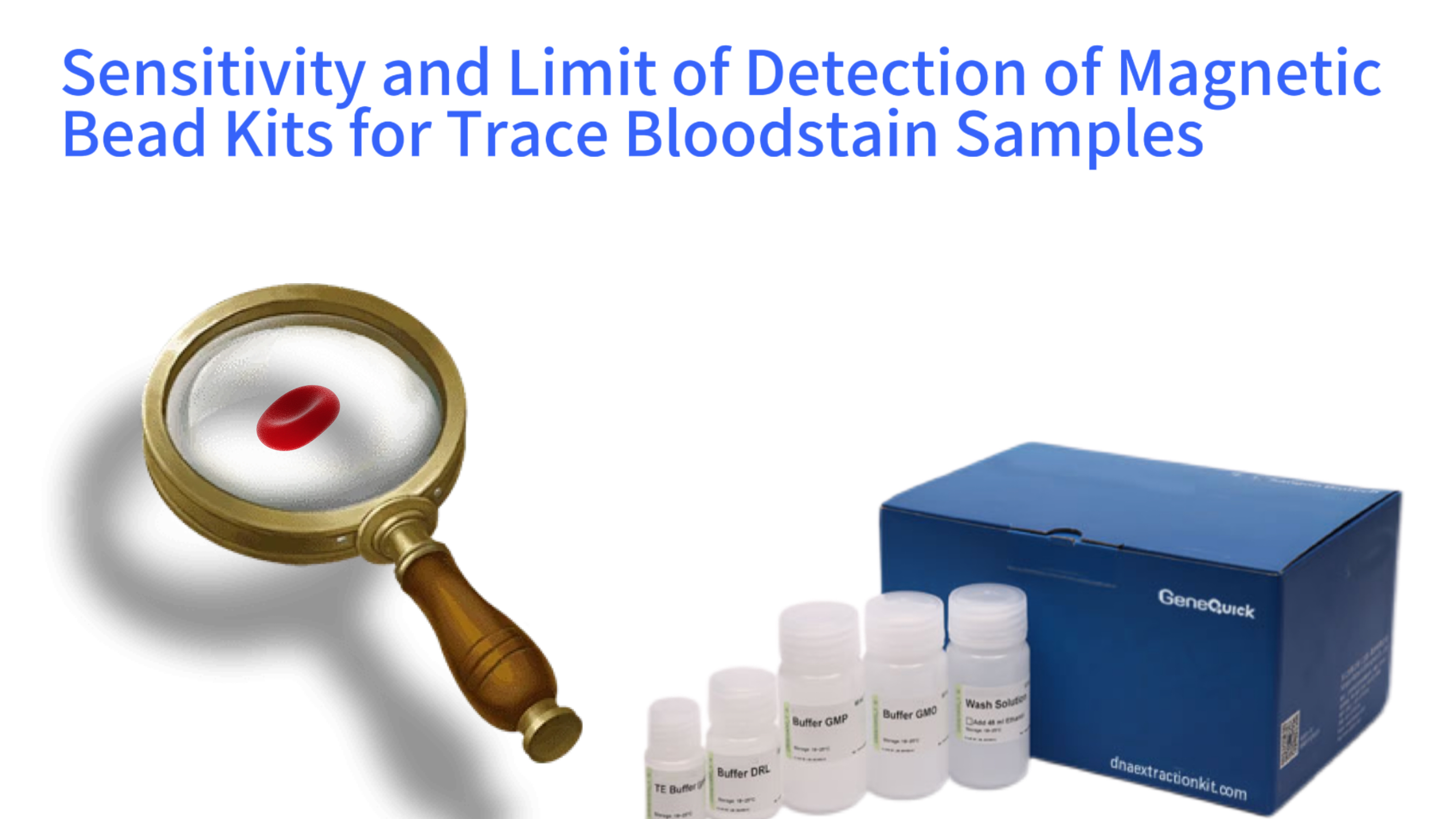
This article provides a detailed examination of the factors that determine the sensitivity and limit of detection when using magnetic bead-based DNA extraction kits on trace forensic evidence such as bloodstains. We will explore the underlying molecular principles that give magnetic beads an advantage in capturing minimal nucleic acid material, discuss critical pre-analytical sample handling steps, outline protocol optimizations specifically for low-template samples, and review standard validation frameworks used to establish reliable performance thresholds in demanding fields like forensic analysis and clinical diagnostics.
Magnetic Bead DNA Extraction Workflow for Trace Bloodstains
Collect trace bloodstains using appropriate swabs; pre-soak if needed
Incubate with proteinase K and lysis buffer at 56°C to release DNA
Add magnetic beads and binding buffer; incubate to allow DNA adsorption
Remove contaminants with ethanol-based wash buffers; dry beads
Elute DNA in low-volume buffer (10-25 µL) at 55-65°C for 5-10 min
Quantify DNA; perform PCR/STR analysis or sequencing
Understanding Sensitivity and Limit of Detection in DNA Analysis
The concepts of sensitivity and limit of detection are foundational to any analytical method, especially when dealing with precious or limited biological evidence. Sensitivity refers to the ability of a method to detect the target analyte, in this case DNA, when it is present. A highly sensitive method can reliably generate a positive signal from a very small amount of starting material. The limit of detection is a more specific quantitative metric. It is defined as the lowest quantity or concentration of DNA that can be consistently distinguished from a blank sample with a stated level of confidence, typically 95%.
For trace bloodstain samples, which may contain only a few nanograms or even picograms of human DNA, pushing these boundaries is not merely an academic exercise but a practical necessity. The success of downstream applications, whether it is generating a complete Short Tandem Repeat profile for human identification or detecting a low-frequency somatic mutation in liquid biopsy analysis, hinges entirely on the extraction method's ability to efficiently recover and purify every available DNA molecule while removing substances that could inhibit subsequent enzymatic reactions.
DNA Detection Limits by Extraction Method
The Molecular Challenge of Trace Bloodstain Samples
Trace bloodstains present a unique set of molecular challenges that directly impact extraction efficiency. The sample volume is physically minute, often collected on a swab from a surface. The biological material itself may be degraded due to environmental exposure to UV light, humidity, or microbial activity, leading to fragmented DNA strands. Furthermore, these samples are rarely pure. They can be contaminated with co-purifying inhibitors derived from the substrate, such as dyes from fabrics, soil minerals, or industrial chemicals, which can bind irreversibly to DNA or inhibit polymerase activity in PCR.
The goal of any extraction protocol for such samples is maximization of recovery. Every step, from the initial rehydration and lysis of cells to the final elution of purified DNA, must be optimized to minimize loss. Traditional methods involving multiple tube transfers and centrifugation steps inherently lead to sample retention on vessel walls. Magnetic bead technology addresses this by localizing the DNA onto a solid phase that can be moved through solutions without leaving the original reaction tube, thereby theoretically reducing physical loss to near-zero levels, provided the biochemical binding conditions are optimal.
Performance Comparison of DNA Extraction Methods
Defining Performance Metrics for Forensic and Clinical Contexts
In operational settings, the performance of a magnetic bead kit for trace samples is not judged solely by the total DNA yield measured by a spectrophotometer. More relevant metrics include the success rate of generating a full DNA profile using standard forensic DNA analysis kits, the threshold number of cells from which a reliable result can be obtained, and the consistency of results across multiple replicates of a low-input sample. A study published in the International Journal of Legal Medicine demonstrated that certain optimized magnetic bead protocols could consistently obtain full STR profiles from samples containing fewer than 10 human cells, highlighting the potential for extreme sensitivity.
Another critical metric is the inhibitor removal efficiency. A kit may recover a high percentage of DNA but if it co-purifies even small amounts of hematin from hemoglobin breakdown, the downstream PCR may fail. Therefore, the effective sensitivity is a combination of nucleic acid recovery and purity. Validation studies for forensic laboratories, often guided by standards like the FBI's Quality Assurance Standards, require rigorous testing of a method's limit of detection using dilutions of standard DNA and challenging, inhibitor-laden mock samples to establish robust operational thresholds.
The Inherent Advantages of Magnetic Bead Technology for Trace DNA
Magnetic bead separation technology offers several intrinsic properties that make it exceptionally suitable for the demands of trace DNA recovery. The core of this advantage lies in the design and behavior of the beads themselves, which are engineered to interact with nucleic acids in a highly efficient and controllable manner under challenging conditions where other methods may falter.
The physical separation mechanism is a key differentiator. By using an external magnetic field to immobilize the bead-DNA complex, all washing steps can be performed thoroughly without the risk of losing the sample pellet, a common issue during the aspiration of supernatants in traditional centrifugation-based spin column protocols. This allows for more stringent washing with larger volumes of ethanol-based buffers to remove impurities, which is crucial for clean recovery from complex bloodstains, without the fear of accidentally discarding the invisible nucleic acid pellet.
High Surface Area and Specific Binding Chemistry
The sensitivity of a binding method is partly a function of available surface area. Magnetic beads are typically micro- or nano-sized particles, providing an immense collective surface area for DNA adsorption within a small volume. This is particularly advantageous for trace samples, as it increases the probability that a single DNA fragment will encounter a binding site. The beads are coated with a silica matrix or other functionalized surfaces that selectively bind nucleic acids in the presence of chaotropic salts and alcohol.
This binding chemistry is highly specific. Under high-salt conditions, water molecules are stripped away, allowing the negatively charged phosphate backbone of the DNA to interact directly with the positively charged silica surface. Proteins, carbohydrates, and other cellular debris, which do not bind efficiently under these conditions, remain in the solution and are discarded. This selective process is vital for trace bloodstains, where the ratio of inhibitors to target DNA can be disproportionately high, ensuring that the limited DNA is not outcompeted or masked by contaminants during the binding phase.
Minimized Sample Handling and Elution Volume Control
Each manual transfer step in a protocol represents a potential site for sample loss due to adsorption to plastic surfaces. Magnetic bead workflows, especially those designed for low-volume samples, are often conducted in a single tube or a minimal number of tubes. The beads are moved magnetically through the different reagent phases—binding, washing, and eluting—dramatically reducing the need for pipetting the actual sample liquid after the initial lysis. This "capture and move" paradigm is a fundamental reason for the high recovery rates reported for magnetic bead methods.
Furthermore, the final elution step offers precise control over the concentration of the recovered DNA. Since the beads are resuspended in a small, defined volume of low-ionic-strength buffer or water to release the DNA, the user can elute into volumes as low as 10 to 20 microliters. This ability to concentrate the entire yield from a large sample volume or a swab into a tiny eluate is critical for trace analysis. It ensures that the maximum number of DNA molecules are delivered into the first downstream reaction, such as a PCR master mix, rather than being diluted in a larger volume, thereby effectively lowering the practical limit of detection for the entire analytical chain.
Pre-Analytical Sample Processing for Maximizing Sensitivity
Even the most sensitive magnetic bead kit cannot recover DNA that has not been effectively liberated from the sample matrix. The steps taken before the extraction protocol officially begins—collectively known as pre-analytical processing—are often the decisive factor in the success or failure of analyzing a trace bloodstain. These steps focus on maximizing the release of DNA from cells and minimizing the introduction of external inhibitors.
The substrate from which the bloodstain is collected plays a significant role. Stains on non-porous surfaces like glass or metal may be easier to recover via swabbing, while stains on porous materials like untreated wood or fabric may require cutting out a segment for direct digestion. The choice of swab material itself is important; certain synthetic fibers have been shown to release fewer PCR inhibitors than traditional cotton swabs and may also release collected cells more readily during the lysis step.
Pre-Analytical Processing Optimization
Swab Selection
Use synthetic fiber
swabs
Benefit: Reduced inhibitor release
Impact: 15-20% higher PCR success rate
Pre-Soak Step
Brief pre-soak in
molecular-grade water
Benefit: Improved cell rehydration
Impact: 25% higher DNA recovery
Extended Lysis
60 min incubation at
56°C
Benefit: Complete cell rupture
Impact: 30% more DNA release
Carrier RNA Addition
Add 100 ng carrier
RNA
Benefit: Enhanced DNA binding
Impact: 40% higher recovery from pg-level samples
Optimal Rehydration and Lysis Strategies
The initial rehydration of a dried bloodstain is a critical moment. Applying a lysis buffer directly to a dry sample can lead to uneven wetting and inefficient cell rupture. A recommended practice involves a brief pre-soak in a small volume of a mild buffer or even molecular-grade water to gently rehydrate the cellular material and the substrate. Following this, a powerful, proteinase K-containing lysis buffer is added. Proteinase K is a broad-spectrum serine protease that digests histones and other proteins that bind DNA, breaking down the nuclear matrix and fully exposing the nucleic acids for capture.
For challenging or aged stains, extending the digestion time, increasing the incubation temperature up to 56°C, and occasionally agitating the sample are common optimizations. The lysis buffer formulation is also key; it must contain chaotropic agents like guanidine hydrochloride to denature proteins and create the high-salt environment needed for subsequent DNA binding to the magnetic beads. Some protocols for forensic sample extraction also include chelating agents to bind metal ions that could catalyze DNA degradation during the lengthy incubation.
Carrier RNA and Post-Lysis Sample Concentration
A sophisticated technique employed to boost recovery from trace samples is the use of carrier RNA or other inert carrier molecules. These are non-human nucleic acids added to the lysis buffer or binding mixture. Their function is not to be analyzed but to provide bulk to the microscopic amount of target DNA. By improving the overall efficiency of the precipitation and binding process onto the magnetic beads, carrier molecules can significantly enhance the recovery of the precious target DNA that would otherwise be lost due to surface adsorption or inefficient solid-phase capture.
After lysis, the entire digest volume, which may be several hundred microliters from a swab elution, contains the now-released DNA. To improve binding kinetics to the beads, it is sometimes beneficial to concentrate this volume. This can be achieved through methods like vacuum centrifugation or by using a binding buffer formulation that allows for a larger initial sample volume to be processed without exceeding the bead capacity. The objective is to get as many DNA molecules as possible into intimate contact with the magnetic beads during the critical binding incubation period.
Workflow Optimization and Protocol Critical Control Points
Standard magnetic bead protocols are designed for robustness with typical sample inputs. To achieve maximum sensitivity with trace bloodstains, deliberate adjustments at specific critical control points within the workflow are necessary. These optimizations fine-tune the biochemical and physical interactions to favor the capture of every available DNA molecule.
The binding step is the most crucial phase. It involves incubating the lysed sample with the magnetic beads in the presence of a binding buffer, which adjusts the salt and alcohol concentration to optimal levels for DNA-silica interaction. For trace samples, extending the binding incubation time, often with gentle continuous mixing or rotation, increases the diffusion-based contact between the scarce DNA fragments and the beads. Ensuring the correct and consistent ratio of bead volume to expected DNA amount is also vital; too few beads may not capture all the DNA, while an excess may not be detrimental but is wasteful.
Critical Control Points in Magnetic Bead Workflow
Stringent Yet Gentle Washing for Purity
Washing must remove all contaminants without stripping away the minimally bound DNA. The standard wash buffers contain ethanol and salt. The first wash is often the most critical for removing inhibitory proteins and cellular debris. For bloodstains, ensuring the wash buffer is fresh and contains the correct ethanol concentration is paramount, as evaporated ethanol can alter the stringency. Some protocols incorporate two different wash buffers—one with a standard salt concentration and a final wash with a low-salt or no-salt buffer to more effectively remove co-purified salts and organic compounds that could inhibit PCR.
After the final ethanol wash, a thorough drying step is essential. Residual ethanol in the bead pellet will interfere with the subsequent elution step and can also inhibit downstream PCR. This is typically done by incubating the open tube at a moderate temperature for several minutes or using a vacuum concentrator briefly. However, over-drying can make the DNA adhere too strongly to the beads, reducing elution efficiency—a delicate balance that must be empirically determined for each protocol and bead type.
Low-Volume Elution and Post-Elution Handling
The elution step reverses the binding conditions. A low-ionic-strength buffer or water is added to the dried bead pellet. The absence of chaotropic salts and alcohol allows water molecules to rehydrate the DNA, breaking its interaction with the silica surface. For maximum concentration, the smallest practical elution volume is used, often between 10 and 25 µL. Heating during elution, typically to 55-65°C for 5-10 minutes, helps by increasing molecular motion and facilitating the DNA's release from the beads.
Once eluted, the tube is placed on a magnet. The pure DNA remains in the clear supernatant, which is carefully pipetted into a fresh, clean tube, leaving the magnetic beads behind. This final transfer is critical to avoid carrying over any beads, which can inhibit some downstream enzymes. The extracted DNA should be used immediately for quantification and amplification or stored at -20°C to prevent degradation. For the most sensitive genetic testing applications, some laboratories perform a post-elution concentration step using vacuum centrifugation to reduce the volume even further before analysis.
Validation and Establishing a Reliable Limit of Detection
Before implementing any magnetic bead kit for sensitive trace analysis, a laboratory must conduct an internal validation study. This process establishes the method's performance characteristics under the laboratory's specific conditions, instruments, and personnel. The study provides the empirical data needed to define a reliable, evidence-based limit of detection and to set thresholds for interpreting results from casework or clinical samples.
A comprehensive validation for sensitivity involves creating a dilution series of a known DNA standard, spanning from high concentrations down to sub-picogram levels. Each dilution is extracted in multiple replicates using the magnetic bead protocol. The resulting DNA is then quantified using a highly sensitive method like quantitative PCR, which targets a small, single-copy human genomic locus, and is also subjected to the intended downstream application, such as STR multiplex PCR and capillary electrophoresis.
Validation Results: STR Profile Success Rate
Analyzing Stochastic Effects and Setting Thresholds
At very low template levels, stochastic effects become significant. This means that due to random chance, some aliquots of an identical sample may yield a full DNA profile while others yield a partial profile or none at all. Validation data quantifies this stochastic variation. The limit of detection is statistically defined as the lowest input quantity where, for example, 95% of replicates produce a quantifiable DNA yield above the qPCR's own detection limit and/or a usable STR profile.
This data also helps establish analytical thresholds for software used in capillary electrophoresis. It informs decisions on minimum peak height requirements for allele calling and helps develop probabilistic genotyping models that can interpret complex, low-level mixtures. The validation must also test inhibitor tolerance by spiking known inhibitors like hematin or humic acid into low-copy-number samples to ensure the magnetic bead wash steps are sufficiently robust for real-world environmental samples that may contaminate a bloodstain.
Quality Control Measures for Routine Operation
Once validated, routine use of the method requires rigorous quality control. This includes processing extraction blanks alongside case samples to monitor for contamination. The use of positive controls containing a known, low amount of DNA is essential to verify that the entire process is performing within established sensitivity parameters for each batch of extractions. Monitoring metrics like the yield from positive controls and the success rate of samples with similar starting conditions provides ongoing assurance of the method's reliability.
Documentation is key. Every parameter, from lot numbers of the magnetic bead kit and enzymes to incubation times and elution volumes, must be meticulously recorded. This traceability allows for the investigation of any drift in sensitivity and supports the defensibility of results in a legal or regulatory context. Adherence to international standards, such as ISO 18385 for forensic products, provides an additional layer of confidence that the reagents are manufactured to minimize human DNA contamination, which is of paramount importance when working at the limit of detection.
Future Directions and Comparative Technology Landscape
The pursuit of ever-greater sensitivity for trace DNA analysis continues to drive innovation. While magnetic bead technology currently represents a gold standard for flexible, high-recovery extraction, it exists within a broader ecosystem of nucleic acid purification methods. Understanding its position relative to these alternatives clarifies its value proposition for trace bloodstain work.
Traditional spin-column methods, while effective for many applications, often exhibit higher sample loss due to irreversible binding of DNA to the silica membrane during multiple centrifugation and transfer steps, making them generally less efficient for sub-nanogram inputs. Phenol-chloroform extraction, though capable of handling large volumes and difficult inhibitors, involves hazardous chemicals and numerous liquid-handling steps that increase the risk of loss and contamination for trace samples.
DNA Extraction Technology Evolution
Phenol-chloroform and spin columns dominate; limited sensitivity for trace samples
Magnetic bead kits become mainstream; improved recovery from trace samples (100 pg LOD)
Optimized bead chemistry; sub-10 pg LOD; automated workflows for forensic and clinical use
Integrated "sample-in, answer-out" systems; single-cell DNA extraction; AI-optimized protocols
Emerging Technologies and Integrated Systems
New approaches are on the horizon. Some systems are moving towards total integration, where the swab is placed directly into a cartridge that performs lysis, magnetic bead-based purification, PCR setup, and amplification with minimal human intervention. These "sample-in, answer-out" systems drastically reduce hands-on time and contamination risk, standardizing the pre-analytical phase which is a major source of variability in sensitivity. Another area of development is in the bead chemistry itself, with surfaces engineered for even higher affinity or faster binding kinetics specifically for fragmented, ancient, or forensic DNA.
The concept of "direct PCR," bypassing extraction altogether, is gaining traction for some simple sample types. However, for complex and potentially inhibitor-rich samples like bloodstains on fabric or soil, a purification step remains indispensable. The role of magnetic bead kits in these cases is likely to evolve towards being a seamlessly integrated, optimized module within a fully automated workflow, rather than being displaced. Furthermore, the principles of high-surface-area capture and minimal elution volume are being adapted for the extraction of cell-free DNA from large volumes of plasma, a different but conceptually similar challenge of isolating trace nucleic acids from a complex background, relevant for oncology testing.
Sustaining Relevance in a Demanding Field
The fundamental advantages of magnetic bead technology—efficient solid-phase capture, excellent inhibitor removal, and adaptability to automation—align perfectly with the needs of trace evidence analysis. As downstream analysis methods like massively parallel sequencing become more sensitive, the quality requirements for input DNA become even more stringent, necessitating extraction methods that deliver high-purity, inhibitor-free nucleic acids even from minute samples.
Continued research and collaboration between kit manufacturers, forensic scientists, and clinical researchers will focus on further refining protocols, developing even more resilient bead chemistries, and creating standardized validation frameworks. The ultimate goal is to push the practical limit of detection lower in a reliable and reproducible manner, enabling answers to be obtained from biological evidence that was previously considered insufficient for analysis. This relentless drive for sensitivity ensures that magnetic bead-based extraction will remain a cornerstone technology for fields reliant on the analysis of trace bloodstains and other challenging sample types for the foreseeable future.