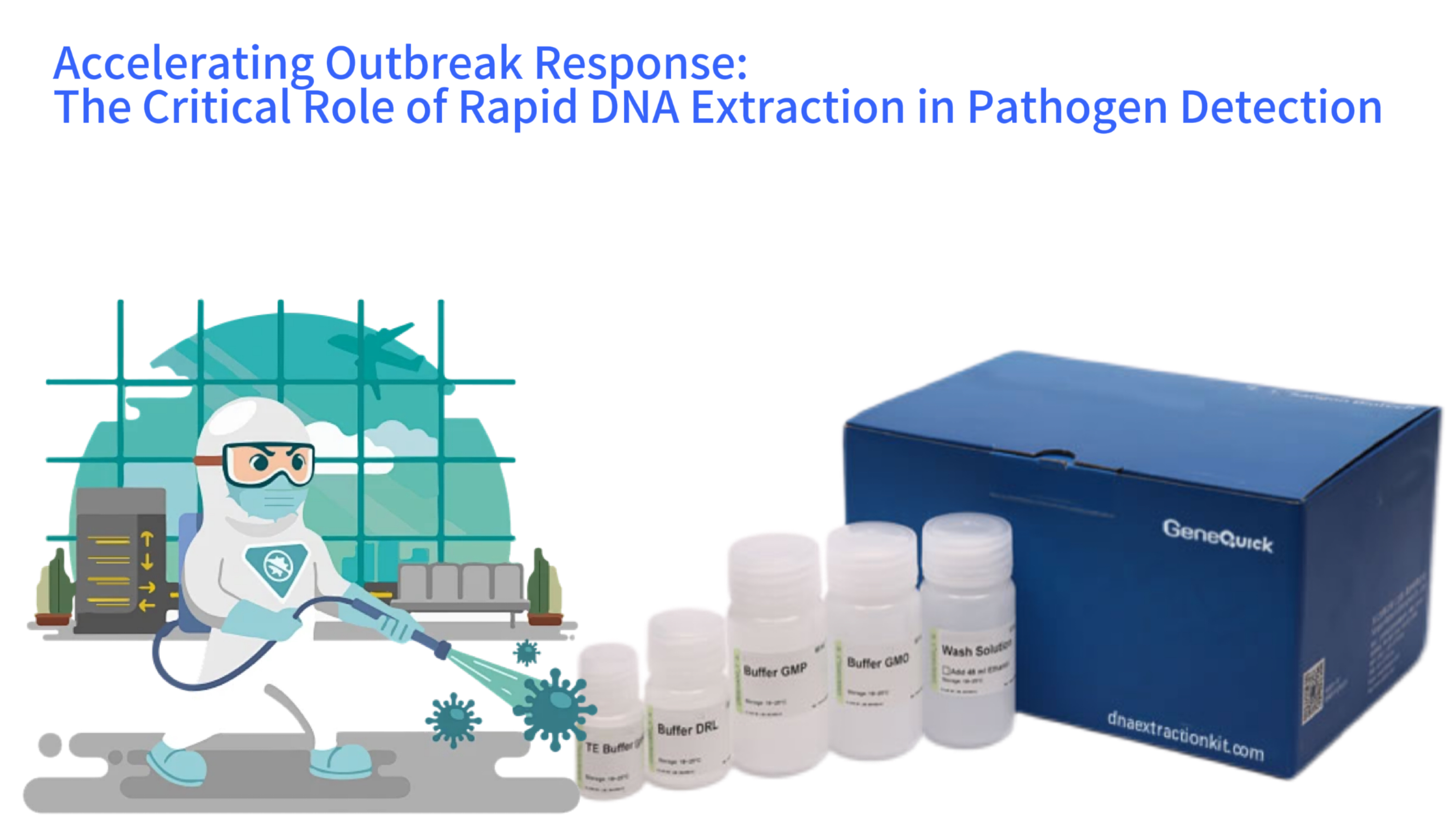
Rapid DNA Extraction & Pathogen Detection Workflow
The emergence of a novel infectious agent or the sudden surge of a known pathogen creates a race against time for public health systems. Identifying the causative agent swiftly is the cornerstone of mounting an effective response, guiding containment measures, and initiating appropriate treatments. At the heart of modern molecular diagnostics lies a critical, yet often overlooked, step: the isolation of pure genetic material from complex clinical samples. Rapid DNA extraction kits have revolutionized this front-line process. This article provides a comprehensive analysis of how these optimized reagent systems are deployed in emergency infectious disease detection. We will examine the technological principles that enable speed without sacrificing purity, detail protocols for handling diverse and challenging emergency samples, and assess the tangible impact these tools have on compressing the timeline from sample collection to actionable result.
The Imperative for Speed in Diagnostic Microbiology
Traditional methods of pathogen identification, such as culture-based techniques, often require days to weeks to yield results. In the context of an outbreak, this delay can be catastrophic, allowing uncontrolled transmission. Molecular methods like polymerase chain reaction (PCR) and next-generation sequencing (NGS) can detect pathogen genetic signatures within hours. However, their success is entirely dependent on the quality of the input nucleic acids. Inhibitors present in clinical matrices like blood, sputum, or stool can completely halt these sensitive reactions. The primary function of a rapid DNA extraction kit in an emergency setting is to deliver inhibitor-free DNA or RNA to the downstream assay in the shortest possible time, transforming a raw sample into a analyzable template with maximum reliability. This process must be robust enough to handle varied, suboptimal, and potentially high-risk samples that are characteristic of outbreak investigations.
The timeline compression offered by rapid kits is significant. A protocol that completes in 20-30 minutes, compared to a multi-hour traditional phenol-chloroform extraction, directly translates to earlier patient diagnosis, quicker cohort isolation, and faster public health notifications. In field laboratories or mobile testing units deployed during outbreaks, the simplicity and minimal equipment requirements of these kits are equally vital. They eliminate the need for hazardous chemicals and complex centrifugation steps, enabling safer and more decentralized testing. The strategic stockpiling of validated rapid extraction kits for key threat pathogens is now a standard component of pandemic preparedness plans for many health agencies globally.
Diagnostic Timeline Comparison
Traditional Methods
Culture-based identification: 2-7 days
Phenol-chloroform extraction: 2+ hours
Total TAT for PCR: 4+ hours
Rapid Extraction Kits
Extraction completion: 20-30 minutes
Total TAT for PCR: ~90 minutes
Time saved per sample: ~210 minutes
Emergency Requirement: Ideal rapid kit workflow<45 minutes (sample input to eluted nucleic acid)
Defining the Requirements for Emergency-Use Extraction
Not all DNA extraction methods are suited for urgent diagnostic scenarios. The ideal rapid kit for emergency pathogen detection must satisfy a stringent set of criteria. Foremost is processing speed, with a complete workflow from sample input to eluted nucleic acid ideally under 45 minutes. This speed must not compromise the two other pillars of performance: yield and purity. The kit must efficiently recover nucleic acid from often low pathogen loads, and it must remove PCR inhibitors common in clinical samples, such as hemoglobin, heparin, ionic detergents, and complex polysaccharides. Furthermore, the protocol must be straightforward, with minimal hands-on time and technical complexity, to reduce human error when laboratory staff are under pressure and processing high volumes.
Another critical requirement is versatility. An outbreak might involve samples from nasal swabs, bronchoalveolar lavage fluid, blood, urine, or even post-mortem tissues. A single kit format that can accommodate this variety without extensive protocol modifications streamlines laboratory operations. Finally, safety and containment are paramount. Protocols that operate in a closed-tube or column-based format minimize aerosol generation, reducing the risk of laboratory-acquired infections when handling dangerous pathogens. The integration of these features—speed, purity, yield, simplicity, versatility, and safety—defines the modern rapid extraction kit for high-stakes diagnostics.
The Impact on Turn-Around-Time and Clinical Decision Making
The integration of rapid DNA extraction directly shortens the total test turn-around-time (TAT). In a typical molecular diagnostic pipeline for a viral infection, the extraction step can constitute a substantial portion of the hands-on and overall processing time. By cutting extraction from 2 hours to 30 minutes, the laboratory can report results a critical 90 minutes sooner. For bacterial sepsis or meningitis, where every hour of delayed appropriate antibiotic therapy increases mortality, this time saving is not merely convenient but lifesaving. Early and accurate identification also allows for the swift discontinuation of broad-spectrum antibiotics in favor of targeted therapy, combating antimicrobial resistance.
Beyond individual patient care, rapid extraction accelerates epidemiological surveillance. During an outbreak, confirming the first cases quickly allows public health teams to define the outbreak's scope, identify the transmission chain, and implement control measures like contact tracing and quarantine more effectively. When combined with high-throughput platforms, rapid extraction kits enable the screening of hundreds of samples per day, providing the data density needed to model outbreak dynamics in real time. This capacity for large-scale, fast testing was a pivotal lesson from recent global pandemics, underscoring the need for scalable and rapid sample preparation solutions at the very start of the diagnostic workflow.
Core Technology: How Rapid Kits Achieve Fast and Pure Nucleic Acid Isolation
The remarkable efficiency of rapid DNA extraction kits stems from a refined and optimized application of the fundamental lysis-binding-wash-elution principle. Manufacturers have engineered each step to minimize incubation periods and manual manipulations while maximizing the selective capture of nucleic acids. The core advancement lies in the formulation of proprietary buffers and the design of the solid-phase binding matrix. These components work in concert to disrupt cells, inactivate nucleases, precipitate contaminants, and create ideal conditions for the instantaneous binding of DNA or RNA to a purification surface, all within a single tube or column. This streamlined biochemistry is what transforms a historically lengthy procedure into a matter of minutes.
Many rapid kits designed for clinical virology or bacteriology also incorporate dual-purpose reagents. For instance, the lysis buffer frequently contains guanidinium salts which simultaneously denature proteins, inactivate most enveloped viruses and bacteria for enhanced laboratory safety, and create a high-salt environment that promotes subsequent binding to silica. This multifunctionality reduces the number of separate reagent additions and incubation steps. The entire process is often amenable to automation, allowing laboratories to link the extraction module directly to liquid handling systems for PCR setup, further reducing hands-on time and potential for cross-contamination in high-throughput emergency testing scenarios.
Rapid DNA Extraction Technology Principle
(Chaotropic salts + detergents)
(Silica membrane/beads)
(Ethanol-based buffers)
(Low-ionic-strength buffer)
Advanced Lysis Formulations for Diverse Pathogens
The initial lysis step must be universally effective against a wide range of microbial cell walls and envelopes. Rapid kits utilize powerful, pre-mixed lysis buffers that are capable of disrupting gram-positive bacteria with their thick peptidoglycan layers, gram-negative bacteria with outer membranes, mycobacteria with waxy mycolic acids, and viruses by solubilizing their lipid envelopes. For some kits targeting intracellular pathogens, an initial proteinase K digestion step may be integrated, but its incubation time is carefully optimized to be just sufficient for tissue or cell disruption without becoming a time bottleneck. The goal is complete nucleic acid release in under 10 minutes.
These buffers are also designed to immediately stabilize the released nucleic acids. Ribonucleases (RNases) and deoxyribonucleases (DNases) are ubiquitous and can degrade the target genetic material in seconds once cells are broken open. The chaotropic salts and detergents in the lysis buffer rapidly denature these enzymes, preserving the integrity of the pathogen's genome for detection. This is especially critical for RNA viruses, whose single-stranded RNA is particularly labile. The stability provided by the lysis chemistry ensures that the genetic signature reaching the PCR assay is representative of the pathogen load in the original sample, a non-negotiable requirement for accurate quantitative diagnostics.
Efficient Binding and Rapid Wash Cycles
Following lysis, the crude mixture contains nucleic acids, proteins, cellular debris, and salts. Rapid kits expedite the separation of target nucleic acids through highly selective binding conditions. In silica-membrane spin columns or magnetic bead systems, the binding chemistry is tuned so that nucleic acids adhere almost instantaneously upon contact with the solid phase under high-salt conditions. This eliminates the need for prolonged binding incubations. The physical design of spin column dna extraction kit units or magnetic separators allows for quick removal of the binding mixture, swiftly moving the process to the wash phase.
The wash steps are equally optimized. Instead of multiple complex buffer changes, rapid kits often employ one or two concentrated wash buffers that efficiently remove salts, proteins, and other contaminants in a single pass. These buffers typically contain ethanol or isopropanol to maintain nucleic acid binding while washing away impurities. Centrifugation times for column-based kits or magnetic separation times for bead-based kits are minimized to just what is necessary for complete buffer exchange. This precision in protocol design shaves critical minutes from each step, culminating in a significantly faster overall process. The final elution uses a low-ionic-strength buffer or nuclease-free water, which releases pure nucleic acids from the binding matrix in a small, concentrated volume ideal for direct use in amplification reactions.
Handling Critical Emergency Sample Types
The chaotic nature of disease outbreaks means diagnostic laboratories receive a heterogeneous array of specimens. A robust rapid extraction protocol must demonstrate consistent performance across these sample types, each presenting unique challenges. The sample matrix itself can be a potent source of PCR inhibitors, and the pathogen may be present in low copy numbers or within complex biological structures. Rapid kits address these challenges through adaptable protocols and buffer systems that can be slightly modified based on the sample input, ensuring that extraction efficiency and purity remain high regardless of the specimen's origin. This adaptability is what makes them indispensable tools in the emergency response toolkit.
Validation studies for clinical rapid extraction kits typically include a panel of the most relevant sample matrices. This validation provides laboratories with verified protocols and expected performance metrics, such as yield and inhibition thresholds, for each sample type. Having this information pre-defined allows laboratory directors to confidently deploy the kit during a crisis without needing to conduct lengthy in-house validation studies. The ability to process swabs, fluids, and tissues with a single core kit system simplifies logistics and training, enabling rapid scaling of testing capacity when it is needed most.
Sample Type Extraction Challenges & Performance
| Sample Type | Key Challenges | Extraction Time | Inhibitor Removal |
|---|---|---|---|
| Respiratory Swabs | VTM stabilizers, low viral load | 20 mins | High |
| Blood/Serum | Heme, IgG, lactoferrin inhibitors | 30 mins | Very High |
| Stool | Bile salts, digestive enzymes | 35 mins | Medium-High |
| FFPE Tissue | Cross-linked DNA, paraffin | 40 mins | High |
Processing Respiratory and Nasopharyngeal Specimens
Swabs from the nasopharynx or oropharynx are frontline samples for respiratory outbreaks, from influenza to novel coronaviruses. These samples are collected in viral transport media (VTM), which preserves pathogen viability but introduces additional proteins and compounds that can inhibit PCR. Rapid extraction kits include protocols specifically for swab/VTM samples, often involving a brief centrifugation or direct aliquot of the VTM into the lysis buffer. The chemistry is designed to overcome common inhibitors found in transport media. The protocol must also efficiently lyse the virus particles to release RNA or DNA, a process made more challenging by the stabilizers in the VTM. Effective kits can handle varying swab types and volumes of VTM, providing a standardized output of pure nucleic acid from what is often a very variable starting material.
For lower respiratory tract samples like sputum or bronchoalveolar lavage (BAL), the challenges include viscosity and the presence of human cellular debris and mucus. Many rapid kits include a pre-treatment step for such samples, such as a dilution with a mucolytic agent or a brief digestion to reduce viscosity. This ensures the sample can be pipetted accurately and that the lysis buffer can penetrate evenly. The binding chemistry must be robust enough to capture pathogen nucleic acids in the presence of high concentrations of competing human DNA. Successful extraction from these samples is critical, as they often contain higher pathogen loads and are key for diagnosing severe pulmonary infections.
Extraction from Blood and Serum for Systemic Infections
Bloodstream infections, whether bacterial, fungal, or viral, represent medical emergencies. Extracting pathogen DNA or RNA directly from whole blood, plasma, or serum is notoriously difficult due to potent PCR inhibitors like heme, immunoglobulin G, and lactoferrin. Rapid kits designed for clinical dna extraction kit for infectious diseases from blood use specialized lysis buffers that precipitate hemoglobin and other proteins while keeping nucleic acids in solution. Some protocols may incorporate a selective step to lyse human blood cells while leaving pathogen cells intact for a subsequent specific lysis, enriching the relative abundance of microbial genetic material.
The small volume of plasma or serum often used in these extractions means the kit must have high binding efficiency to recover low-copy-number targets, which is common in early-stage bacteremia or viremia. For sepsis diagnosis, the speed of the extraction directly influences the time to species identification and antimicrobial resistance gene detection. Modern rapid kits for blood samples can often process specimens in under 30 minutes, delivering template suitable for real-time PCR or multiplex panels that can identify a broad panel of pathogens and resistance markers from a single extraction, a powerful combination for guiding life-saving therapy.
Managing Complex and Challenging Matrices
Outbreak investigations can extend to unconventional samples. Stool specimens are crucial for diagnosing enteric pathogens like norovirus, Salmonella, or E. coli, but they contain complex mixtures of bacteria, digestive enzymes, and bile salts that are severe PCR inhibitors. Rapid kits for stool include strong inhibitors-removal steps, sometimes involving a separate inhibitor-removal resin or bead in addition to the standard binding matrix. The protocol is designed to separate the inhibitor-laden supernatant from the nucleic acid-bound solid phase with extreme thoroughness during washing.
Similarly, tissue samples from biopsies or autopsies may be formalin-fixed and paraffin-embedded (FFPE), which cross-links and fragments nucleic acids. While not always a "rapid" process due to the necessary deparaffinization steps, specialized rapid FFPE kits optimize the protease digestion and reversal of cross-links to shorten the time to extract usable DNA from these valuable archival samples during outbreak trace-back investigations. For all complex matrices, the magnetic beads dna extraction kit technology offers advantages, as magnetic separation can be more effective than centrifugation in removing fine particulate inhibitors that might clog a spin column membrane.
Technology Selection: Comparing Platforms for Emergency Deployment
Laboratories preparing for outbreak response must choose a rapid extraction platform that aligns with their anticipated testing volume, available equipment, and required sample types. The two dominant technologies are silica-membrane spin columns and magnetic bead-based systems, each with distinct operational profiles. Spin column kits are widely used, familiar to most technologists, and require only a microcentrifuge and vortex. They are excellent for low-to-mid throughput and offer consistent, high-purity yields. Their process is largely manual, which can become a bottleneck when processing hundreds of samples during a surge.
Magnetic bead systems offer greater potential for automation and higher throughput. The bead-nucleic acid complexes are separated using a magnet, eliminating centrifugation steps. This allows the process to be easily adapted to robotic liquid handlers, enabling walk-away automation for 96-well plates or more. This is a decisive advantage in large reference laboratories or during a massive outbreak. The choice between these platforms often comes down to a trade-off between upfront simplicity and long-term scalability for surge capacity. Some laboratories maintain both, using columns for stat samples and batched bead-based extraction for surveillance testing.
Extraction Platform Comparison for Emergency Use
Silica-Membrane Spin Columns
Equipment Requirement: Microcentrifuge only
Throughput: Low-mid (manual processing)
Purity: High (A260/A280 ~1.8)
Time per sample: 20-30 mins
Best For: Field labs, low-volume testing, resource-limited settings
Magnetic Bead Systems
Equipment Requirement: Magnetic stand/automated workstation
Throughput: High (96-well automation)
Purity: High (A260/A280 ~1.7-1.8)
Time per batch: 30-40 mins (96 samples)
Best For: Large outbreaks, high-volume screening, reference labs
Spin Column Kits: Reliability and Accessibility
Silica-membrane spin columns represent a mature and highly reliable technology. The user mixes the lysed sample with a binding solution and applies it to a column containing a small silica filter. During a brief centrifugation, nucleic acids bind to the silica membrane while contaminants pass through. Subsequent wash and elution steps are performed with further centrifugations. The entire process is visually intuitive and contained within the column assembly, minimizing spill risk. The quality of DNA from these kits is typically very high, with excellent A260/A280 ratios indicating low protein contamination.
For emergency settings in resource-limited areas or field laboratories, the minimal equipment requirement of a microcentrifuge makes spin column kits particularly accessible. They do not require electricity for magnetic stands or complex robotics. Newer versions of these kits have dramatically reduced centrifugation times—sometimes to 1 minute or less per spin—placing them firmly in the "rapid" category. Their main limitation is manual throughput; processing a large batch of 96 samples can be labor-intensive and time-consuming due to the sequential handling of individual columns or strips of 8 or 12.
Magnetic Bead Systems: Throughput and Automation Potential
Magnetic bead-based extraction operates on a similar binding principle but uses paramagnetic silica beads as the solid phase. After binding in a tube or plate well, a magnet is applied to the side or bottom of the vessel, pulling the bead-nucleic acid complex to the wall and immobilizing it. The liquid supernatant containing impurities can then be simply aspirated and discarded. Wash buffers are added and removed in the same magnetic separation manner, followed by elution. This "capture and release" mechanism without centrifugation enables true high-throughput processing.
The automation compatibility of magnetic systems is their most powerful feature for emergency response. Automated workstations can process dozens of samples in parallel with minimal human intervention, standardizing the process and freeing skilled staff for result analysis and reporting. This scalability is essential for community-wide screening or testing in a major outbreak. Furthermore, magnetic separation can be gentler on long genomic DNA fragments, which is beneficial for techniques like whole-genome sequencing used to track transmission chains. The trade-off is the need for a higher initial investment in magnets or automated platforms and slightly more technical optimization of the liquid handling steps in the protocol.
Quality Control and Contamination Prevention in High-Pressure Settings
When operating at high speed and volume during an outbreak, the risks of procedural errors and cross-contamination between samples increase. A false-positive result due to contamination can misdirect public health resources and cause unnecessary public alarm. Therefore, the design of rapid extraction kits and the associated laboratory workflow must incorporate robust safeguards. Many kits include built-in negative extraction controls and recommend the use of external controls. The physical design of the kits themselves, such as pre-aliquoted reagents and single-use tips or columns, is a first line of defense against carry-over contamination.
Laboratories must establish strict unidirectional workflow practices, physically separating pre-PCR (extraction) areas from post-PCR (amplification and detection) areas. The use of dedicated equipment, lab coats, and consumables for each area is non-negotiable. Within the extraction process, the inclusion of a DNase or RNase treatment step in certain kits can help degrade contaminating amplicons from previous PCR runs, a common source of false positives. Adherence to international standards for molecular testing, such as those outlined by organizations like the College of American Pathologists (CAP) or ISO, provides a framework for maintaining quality even under the duress of an emergency response.
Quality Control Framework for Rapid Extraction
Integrating Internal and External Controls
Every batch of extractions performed during outbreak testing should include control samples to monitor kit performance and laboratory technique. An external positive control, containing a known quantity of non-infectious synthetic or inactivated pathogen material, is extracted alongside patient samples. Its successful detection confirms that the extraction and the downstream PCR assay functioned correctly. An external negative control, such as nuclease-free water or negative matrix, is processed identically to check for reagent contamination.
Some advanced rapid extraction kits incorporate an internal control (IC) directly into the lysis buffer. This IC is a non-pathogenic nucleic acid sequence that is spiked into every sample at the beginning of extraction. It co-extracts and co-amplifies with the target pathogen sequence. Detection of the IC in the final PCR assay confirms that the individual sample extraction was successful and that no significant inhibitors were carried over. Failure of the IC signal indicates a problem with that specific sample's extraction, alerting the technologist to a potential false-negative result and the need for re-testing. This sample-specific quality check is invaluable for maintaining confidence in negative results during high-stakes testing.
Workflow Design to Mitigate Amplicon Contamination
The most pernicious form of contamination in a molecular laboratory is amplicon contamination—the carry-over of PCR products from previous amplification reactions into new samples. These amplicons can serve as potent templates in subsequent PCRs, generating false-positive signals. Preventing this requires meticulous laboratory practice. The use of rapid dna extraction kit systems that are largely closed-tube, where reagents are added without exposing the interior to the environment, is beneficial. Many automated bead-based systems perform all liquid transfers with sealed tips, offering a high degree of containment.
Physical separation remains the gold standard. Extraction and PCR setup should be performed in a dedicated clean room or cabinet with positive air pressure, while amplification and detection occur in a separate room. Reagents and equipment should never move from the post-PCR area back to the pre-PCR area. The incorporation of uracil-DNA glycosylase (UDG) into master mixes is a common biochemical safeguard; it degrades any contaminating amplicons from previous runs that contain uracil before the new PCR begins. Together, these layered strategies ensure that the speed of rapid extraction does not come at the cost of result accuracy.
A Decision Framework for Implementing Rapid Extraction in Emergency Preparedness
Selecting and implementing the right rapid DNA extraction solution for an institution's outbreak response plan requires a strategic assessment. The decision should be guided by a clear understanding of the expected threat pathogens, the types and volumes of samples anticipated, existing laboratory infrastructure, and available staff expertise. A preparedness plan that includes pre-validated rapid extraction protocols for priority pathogens allows a laboratory to transition to emergency operations seamlessly. This involves not just purchasing kits, but also training staff, establishing workflows, and ensuring a sustainable supply chain for critical consumables.
The framework should also consider future scalability. A system that works well for the sporadic cases of a routine season may be overwhelmed during an epidemic. Partnerships with manufacturers for guaranteed supply during a crisis, or the identification of alternative but validated kit options, are prudent steps. Furthermore, the chosen technology should be compatible with the laboratory's downstream detection platforms, whether they are end-point PCR, real-time PCR, or NGS. The goal is to create an integrated, resilient diagnostic pipeline where rapid extraction is the reliable first act, setting the stage for accurate and timely pathogen identification.
Emergency Preparedness Decision Framework
Step 1: Needs Assessment
- Identify priority pathogens (respiratory, bloodstream, enteric)
- Define sample types and expected volume
- Evaluate
existing infrastructure (equipment, staff)
Step 2: Technology Selection
- Spin columns: Low-volume/field use
- Magnetic beads: High-volume/outbreak response
- Validate compatibility with downstream
assays (PCR/NGS)
Step 3: Operational Resilience
- Stockpile kits and ancillary
supplies
- Train multiple staff on protocols
- Establish supply chain backups
- Integrate with LIMS for rapid result
reporting
Assessing Needs Based on Pathogen and Sample Profile
The first question in the selection process is: what are we preparing for? A public health laboratory may prioritize kits validated for respiratory viruses and vector-borne diseases, while a hospital lab may focus on bloodstream and central nervous system infection panels. The sample types dictate protocol requirements; a lab that primarily receives swabs will have different needs than one processing sputum or tissue. The expected pathogen load also matters. For low-load targets like latent viruses or early HIV infection, a kit with exceptionally high yield and low inhibition is necessary, even if it is slightly slower. For high-load outbreaks, speed and throughput may be the dominant factors.
It is also essential to consider the nucleic acid type. While many kits are designed for total nucleic acid extraction, some outbreaks are caused exclusively by RNA viruses. In such cases, a kit optimized for RNA recovery and stability, potentially with an integrated DNase step to remove background human DNA, might offer performance advantages. Consulting published validation studies from public health agencies for specific pathogen-kit combinations provides evidence-based guidance for this critical selection. For example, during a viral hemorrhagic fever outbreak, using a kit with a proven track record for safety and efficiency with that specific virus family in forensic dna extraction kit for blood or serum samples would be a prioritized choice.
Evaluating Infrastructure and Building Operational Resilience
The choice between spin column and magnetic bead technologies hinges on the laboratory's existing equipment and capacity for investment. A lab with several microcentrifuges and trained staff in manual protocols may opt to scale up by stockpiling column-based kits and implementing shift work. A lab planning for high-volume testing might invest in a magnetic bead extraction platform and a liquid handling robot, accepting a higher upfront cost for vastly greater surge capacity and reduced labor burden during a crisis.
Operational resilience extends beyond the kit itself. It includes ensuring an uninterrupted supply of essential ancillary items: sterile pipette tips, PCR tubes, microcentrifuges in good repair, and reliable cold storage for reagents. Training programs should ensure that more than one technologist is proficient in the extraction protocol. Finally, the laboratory's information management system must be able to handle a sudden influx of samples and track them from receipt through extraction, PCR, and result reporting without error. A rapid extraction is only valuable if the result reaches the clinician or public health official quickly and accurately; the entire system must be designed for speed and reliability.