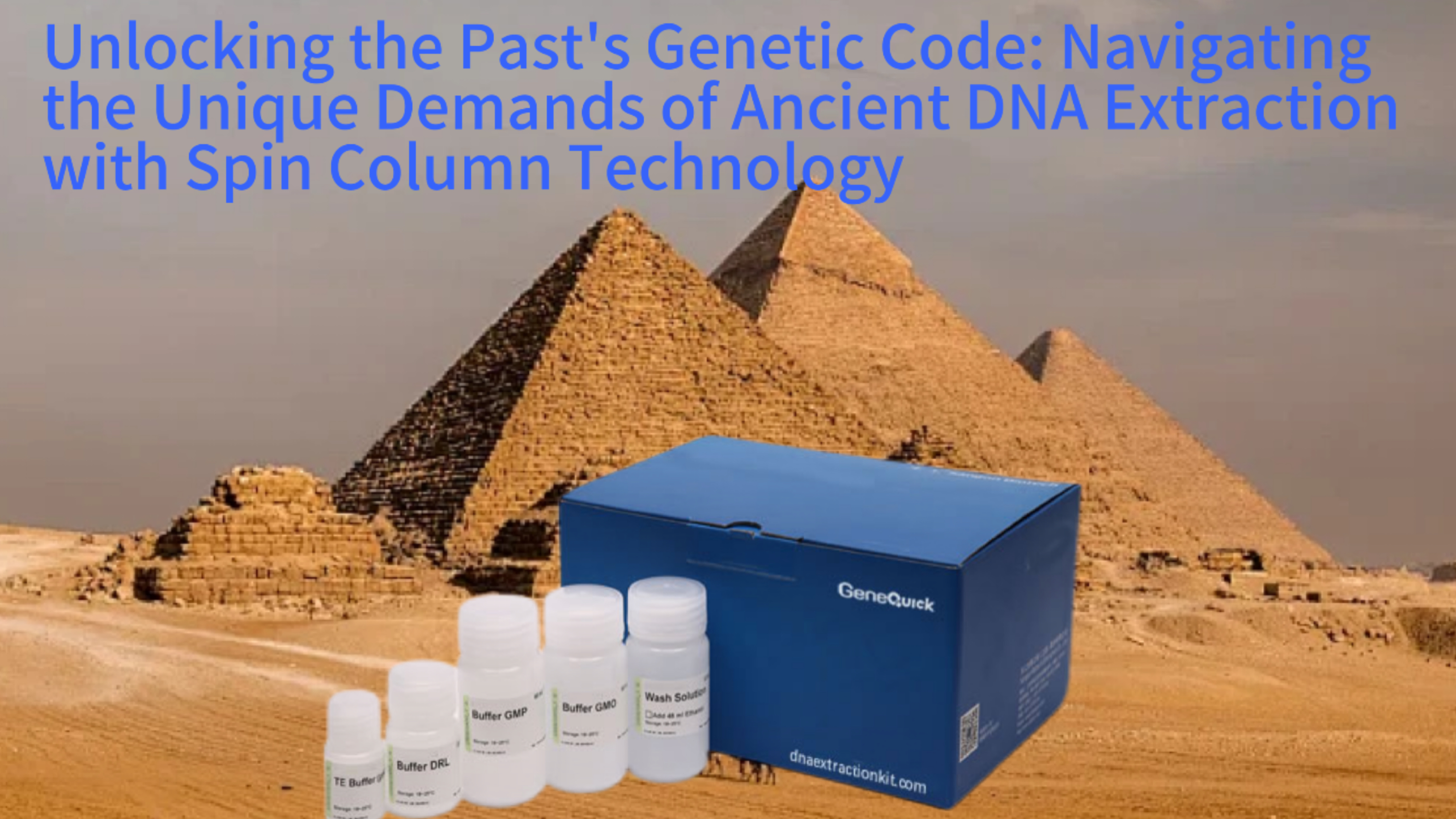
The quest to recover and sequence DNA from ancient specimens, such as bones, teeth, and preserved tissues, represents one of the most challenging frontiers in molecular biology. Ancient DNA (aDNA) research offers unparalleled insights into evolutionary history, population migrations, and extinct species, but the very nature of the sample material presents a formidable obstacle. The DNA within these specimens is not merely old; it is profoundly degraded, fragmented into short pieces often fewer than 100 base pairs in length, and exists in minute quantities amidst a background of overwhelming environmental contamination and PCR inhibitors. This extreme context elevates the choice of extraction methodology from a routine procedural step to a critical determinant of experimental success or failure. The spin column DNA extraction kit, a cornerstone of modern molecular laboratories due to its simplicity and reliability, must be carefully evaluated and adapted for this specialized field. While its core principles of silica-based binding remain valid, its application in aDNA work is defined by a unique set of challenges centered on maximizing the recovery of ultrashort, damaged molecules while minimizing the introduction of modern contaminants. This analysis explores the specialized protocols, inherent limitations, and critical adaptations required to deploy spin column technology effectively in the rigorous and delicate world of ancient DNA research.
The Inherent Nature of Ancient DNA: A Sample in Crisis
Ancient DNA is not simply a dilute version of modern DNA. It is a molecular product of extensive post-mortem damage processes that fundamentally alter its physical and chemical state. Over centuries or millennia, hydrolytic and oxidative reactions systematically break the phosphate backbone of the DNA molecule. This results in extreme fragmentation, with the average fragment length decreasing exponentially over time. In specimens tens of thousands of years old, the majority of recoverable DNA may be shorter than 50 base pairs. Concurrently, base modifications occur, with deamination of cytosine to uracil being a particularly common lesion. This damage creates apparent C-to-T transitions during sequencing, which must be bioinformatically distinguished from true genetic variants.
Beyond molecular damage, the sample matrix itself is hostile. Ancient remains, especially bones and teeth acting as a partial mineral shield, often contain co-purifying substances that are potent inhibitors of downstream enzymatic reactions like PCR. These include humic acids from soil, collagen breakdown products, and calcium ions from the mineral matrix. Furthermore, the original endogenous DNA from the target organism is typically outnumbered by DNA from environmental microbes and, most problematically, modern human DNA introduced during excavation or handling. The initial quantity of endogenous aDNA can be vanishingly small, sometimes as low as a few picograms, making every step in the extraction process a potential point of catastrophic loss. This context defines the primary objectives for any extraction method: to selectively capture the shortest fragments, to aggressively remove inhibitors, and to operate within an ultra-clean, contamination-controlled workflow.
Post-Mortem Degradation: Fragmentation and Chemical Damage
The fragmentation of aDNA is a continuous process driven by hydrolysis. Water molecules attack the phosphodiester bonds that form the backbone of the DNA double helix. In porous substrates like bone, this process is accelerated. The result is a population of molecules of random lengths, heavily skewed toward the very short. This physical degradation has direct implications for extraction efficiency. Many standard DNA extraction protocols, including some spin column designs, have binding and washing conditions optimized for longer, modern DNA. They may fail to retain these ultrashort fragments or may wash them away during the purification steps, leading to a complete loss of the already scarce genetic signal.
Chemical damage further complicates the picture. Deamination of cytosines into uracils, which base-pairs like thymine, is a time-dependent process. In double-stranded regions, this damage often occurs at the single-stranded overhangs of fragments. These lesions are not just sequencing artifacts; they can interfere with adapter ligation in next-generation sequencing library preparation. The extraction process itself must be gentle enough to not exacerbate this damage. Harsh lysis conditions involving excessive heat or vigorous physical disruption can accelerate strand breakage in already fragile molecules. Therefore, the initial sample digestion for aDNA often employs prolonged, low-temperature incubation with proteinase K in a neutral buffer, a stark contrast to the rapid, high-temperature lyses used for fresh tissue.
The Challenge of Contaminants: Inhibitors and Microbial DNA
The environment surrounding an ancient specimen is a rich source of confounding biomolecules. Soil-derived humic and fulvic acids are polyphenolic compounds that co-extract with DNA and are powerful inhibitors of Taq polymerase. They can bind to the DNA itself or directly inactivate enzymes, leading to PCR failure even when DNA is present. Standard spin column wash buffers, often based on ethanol or isopropanol with mild salts, are frequently insufficient to remove these stubborn contaminants. Their presence necessitates additional wash steps or the use of specialized wash buffers containing guanidine salts or other chaotropic agents at different pH levels to disrupt their interaction with the silica membrane or the DNA molecule.
Perhaps an even greater challenge is the sheer abundance of non-target DNA. Microbial communities colonize the specimen after death, and their intact, modern DNA is often orders of magnitude more abundant than the degraded endogenous host DNA. While spin columns bind DNA largely irrespective of source, this means the final eluate contains a complex mixture. For studies targeting the host genome, this microbial DNA constitutes background noise that consumes sequencing depth. For paleomicrobiology studies aiming to characterize ancient microbes, the challenge is to distinguish ancient microbial sequences from those of modern contaminants that infiltrated the sample post-excavation. This differentiation is not achieved during extraction but relies on downstream computational methods and strict laboratory controls.
Low Endogenous DNA Content and Modern Contamination Risk
The absolute quantity of authentic ancient DNA in a sample extract is frequently minuscule, existing at the very limits of detection. This scarcity amplifies the risk of contamination by modern DNA, which is ubiquitous in laboratories and on researchers. A single skin cell shed during handling can contain thousands of copies of modern human DNA, completely overwhelming the signal from an ancient human bone. Therefore, the physical process of extraction must be conducted in a dedicated, physically isolated ancient DNA laboratory with positive air pressure, UV irradiation, and stringent personnel protocols, including full-body suits and face masks.
The spin column kit itself becomes a potential vector for contamination. Reagents must be certified nuclease-free and, ideally, tested for the absence of human DNA. The use of liquid handling robots, while reducing human contact, must be carefully validated to ensure they do not carry over contamination between samples. The entire workflow, from powdering the bone to the final elution, is designed as a one-way process from the clean lab to the post-PCR area. This environmental control is not a feature of the kit but a mandatory framework within which the kit must operate. The choice of a spin column system that minimizes open-tube steps and utilizes sealed, single-use columns can support, but never replace, these overarching sterile techniques.
The Spin Column Mechanism: Suitability and Limitations for aDNA
The fundamental principle of spin column DNA extraction is the selective binding of nucleic acids to a silica-based membrane in the presence of high concentrations of chaotropic salts, such as guanidine hydrochloride or thiocyanate. These salts disrupt the hydration shell around the DNA molecule, allowing the negatively charged phosphate backbone to interact with the positively charged silica surface. After binding, impurities like proteins, salts, and cellular debris are washed away with ethanol-based buffers. Finally, the purified DNA is eluted in a low-ionic-strength buffer or water, which rehydrates the molecule and disrupts its interaction with the silica. This process is highly effective for clean, high-molecular-weight DNA but requires critical evaluation for aDNA applications.
For ancient DNA, the primary advantage of spin columns is their ability to produce inhibitor-free eluates suitable for sensitive downstream applications like PCR and sequencing. The multi-step wash process can be highly effective at removing humic acids and other PCR inhibitors when optimized. Furthermore, spin column protocols are easily scalable and automatable, processing multiple samples in parallel. However, the technology has inherent drawbacks for ultrashort fragments. The binding kinetics favor longer molecules, and the centrifugal force and wash steps can lead to disproportionate loss of the shortest fragments. Additionally, the silica membrane has a finite binding capacity, which is rarely a concern for modern DNA but can become saturated with the vast excess of environmental microbial DNA in ancient extracts, reducing the already low yield of endogenous material.
Silica Binding Kinetics and Fragment Size Bias
The interaction between DNA and silica is influenced by the length of the DNA molecule. Longer molecules have more phosphate groups to interact with the silica surface, leading to stronger, multivalent binding. Very short fragments, with only a few dozen base pairs, have fewer points of contact. This makes their binding less efficient and more reversible. During the subsequent wash steps, where buffers are passed through the column by centrifugation, these weakly bound short fragments are more likely to be dislodged and lost. This creates a systematic bias in the extracted DNA population, enriching for longer molecules that may represent modern contamination or better-preserved microbial DNA, while depleting the most degraded, ancient endogenous sequences.
To mitigate this bias, protocols for aDNA extraction using spin columns often modify the standard binding conditions. Increasing the volume or concentration of the binding buffer containing chaotropic salts can improve the capture of short fragments. Some protocols also incorporate carrier molecules, such as poly-A RNA or glycogen, which do not interfere with downstream analyses but help precipitate and bind short DNA fragments by providing a "backbone" that enhances silica interaction. However, the addition of carriers must be carefully considered, as they can also co-precipitate inhibitors or increase background in sensitive assays. The goal is to shift the binding equilibrium to favor the retention of the precious, fragmented aDNA without compromising the purity of the final extract.
Wash Stringency and Inhibitor Removal Efficiency
The wash steps in a spin column protocol serve a dual purpose: removing salts and chaotropic agents from the binding step and eliminating non-specifically bound organic and inorganic impurities. For ancient samples, the composition and stringency of these washes are paramount. Standard washes with 70-80% ethanol effectively remove salts and alcohol-soluble contaminants but may be inadequate for the polyphenolic inhibitors common in archaeological specimens. Consequently, specialized aDNA protocols often incorporate an additional wash with a buffer containing guanidine salt at a different pH or a proprietary detergent blend designed to solubilize humic substances.
The challenge lies in balancing purity with yield. Increasing wash stringency by using larger volumes, higher alcohol concentrations, or additional wash steps improves purity by more thoroughly removing inhibitors. However, each additional centrifugation step also increases the mechanical shearing force on the bound DNA and the risk of losing the shortest, most weakly bound fragments. Over-washing can result in a perfectly clean eluate that contains almost no DNA. Therefore, protocols must be empirically optimized for specific sample types, such as bone versus tooth dentine versus sediment, to find the precise wash conditions that maximize inhibitor removal while minimizing aDNA loss. This optimization is a form of specialized method development unique to the ancient DNA field.
Elution Considerations: Volume, Temperature, and Efficiency
The final elution step aims to recover the bound DNA in a minimal volume to achieve a high concentration, which is critical for subsequent library preparation. Standard elution uses a low-ionic-strength Tris-EDTA buffer or nuclease-free water, often heated to 55-70°C to increase solubility and detachment from the silica membrane. For ancient DNA, this step requires careful consideration. Using too small an elution volume risks leaving a significant portion of the DNA bound to the column, as the diffusion of molecules out of the silica matrix is not perfectly efficient. A larger elution volume ensures higher recovery but yields a more dilute extract, which may require a concentration step that risks further loss.
Furthermore, the elevated temperature used to promote elution could potentially accelerate hydrolysis of the already damaged DNA strands, though brief incubation is generally considered safe. Some protocols recommend a two-step elution: applying a first elution buffer, incubating, spinning it down, and then applying a second aliquot of buffer to the same column to recover residual DNA. This can increase total yield by 15-30%. The eluate is often not used directly but is concentrated using centrifugal vacuum concentrators or by performing a secondary binding and elution into a smaller volume. Each of these manipulations introduces additional opportunities for contamination and sample loss, underscoring the delicate trade-offs in every stage of aDNA extraction with spin columns.
Specialized Protocol Adaptations for Ancient Samples
To address the unique challenges of ancient DNA, researchers have developed extensive modifications to standard commercial spin column kit protocols. These adaptations are not mere suggestions but essential revisions born from empirical testing across thousands of samples. The workflow begins long before the kit is opened, with stringent decontamination of the sample surface, often through physical abrasion, bleach washes, or UV irradiation, to remove exogenous contamination. The sample is then converted into a powder, typically using a dedicated drill in a clean lab environment, to maximize surface area for digestion.
The lysis step is profoundly different. Instead of a quick, high-temperature incubation, ancient bone or tooth powder is digested for 12 to 24 hours at 37-56°C in a buffer containing EDTA to decalcify the mineral matrix and proteinase K to degrade the collagenous and cellular structures. This long, gentle digestion is crucial to fully release the DNA without causing further fragmentation. After digestion, the supernatant is carefully separated from the insoluble pellet, often through centrifugation. It is at this point that the lysate is combined with the binding buffer from the spin column kit, initiating the silica-based purification phase. However, even this transfer is optimized, with some protocols including a pre-treatment step to remove particularly stubborn inhibitors before binding.
Pre-Binding Treatments and Decalcification Strategies
For skeletal elements, the first major hurdle is decalcification. The hydroxyapatite mineral matrix of bone and tooth traps DNA and must be dissolved to release it. This is achieved by incubating the powdered sample in a high-concentration EDTA buffer, which chelates calcium ions. This step can take several hours to overnight. The efficiency of decalcification directly impacts DNA yield. Incomplete decalcification leaves a significant portion of the DNA inaccessible within the mineral lattice. Following decalcification, proteinase K is added to digest the organic component, primarily collagen. The long incubation times ensure that even highly cross-linked proteins are broken down, liberating their bound DNA.
Before the lysate is added to the spin column binding buffer, it may undergo a pre-cleaning step. One common method is the use of a binding buffer without silica, where the lysate is mixed with chaotropic salts and then centrifuged to pellet insoluble inhibitors and particulates. The supernatant, now partially clarified, is then transferred to a fresh tube for the actual binding to the silica column. Another strategy involves size-selective precipitation or the use of specific resins to absorb humic acids. These pre-treatments add complexity but can dramatically improve the purity of the DNA that ultimately reaches the spin column, enhancing both binding efficiency and the quality of the final eluate. Similar meticulous pre-treatment logic applies to other complex matrices, as seen in protocols for environmental soil samples.
Modified Binding and Incubation Steps
Once the clarified lysate is ready, the binding step is carefully controlled. The standard protocol of adding an equal volume of binding buffer is followed, but the mixture is often incubated on ice or at room temperature for an extended period, sometimes 10-30 minutes, before being loaded onto the column. This extended incubation time allows the chaotropic salts to fully denature proteins and disrupt inhibitor complexes, and it gives the short aDNA fragments more time to interact with the binding solution, potentially improving their capture efficiency. The lysate-binding buffer mixture is then loaded onto the silica membrane slowly to avoid overloading and to ensure optimal contact time.
After loading, the column is not immediately centrifuged. A second incubation period of several minutes is common, allowing the DNA to bind to the silica membrane under static conditions. This minimizes the immediate shearing force that centrifugation would apply, which could dislodge short fragments. Only after this pause is the column centrifuged at a moderate, controlled speed. Some protocols even split the lysate across multiple columns to avoid exceeding the binding capacity of a single membrane, ensuring that every available molecule of endogenous DNA has a binding site and is not outcompeted by the more abundant environmental DNA. These meticulous timing and volume adjustments transform a routine step into a critical recovery phase.
Enhanced Wash Regimens and Final Recovery
The wash phase is where protocols diverge most significantly from kit instructions. A standard two-wash ethanol protocol is almost never sufficient. A typical aDNA-optimized wash regimen might include: 1) A first wash with the kit's standard wash buffer (often guanidine-based) to remove bulk contaminants. 2) A second, more stringent wash with a buffer containing a higher percentage of ethanol or a different chaotropic salt formulation to remove residual organics. 3) A final "polishing" wash with 80% ethanol to remove all traces of salts and chaotropic agents. Between washes, columns are centrifuged thoroughly to remove all liquid, and the flow-through is discarded from the collection tube to prevent carryover.
After the final wash, an additional "dry spin" with the column empty is performed to evaporate residual ethanol, which can inhibit elution and downstream enzymatic reactions. For elution, pre-warmed elution buffer is applied directly to the center of the silica membrane, not to the column walls. The column is then incubated at room temperature for 5 minutes before centrifugation. To maximize recovery, this elution step is frequently repeated: the first eluate is collected, a fresh aliquot of buffer is added to the same column, incubated again, and centrifuged. The two eluates are combined, representing the total yield. This final extract is then quantified using fluorometry methods sensitive to double-stranded DNA, though the results often reflect total DNA (including microbial) rather than endogenous aDNA. The purified DNA is now ready for conversion into sequencing libraries, a process that itself has been revolutionized to accommodate short, damaged fragments. The entire workflow, from powder to library, embodies the specialized nature of working with degraded biological evidence.
Downstream Application Compatibility and Quality Assessment
The ultimate validation of any ancient DNA extraction is its performance in downstream molecular applications. The extracted DNA must serve as a viable substrate for enzymatic reactions, primarily PCR for targeted analyses or next-generation sequencing (NGS) library preparation for genome-wide studies. The presence of inhibitors, even at trace levels undetectable by spectrometry, can cause complete failure of these sensitive steps. Therefore, quality assessment for aDNA extracts focuses not just on quantity but, more critically, on functionality and authenticity.
Standard metrics like the A260/A280 and A260/A230 ratios from a spectrophotometer are of limited utility for aDNA. The very low concentrations often fall below the reliable detection limit of these instruments, and the ratios can be skewed by the presence of co-purified RNA, proteins, or specific inhibitors that absorb at 230 nm. More informative is fluorescence-based quantification using dyes like PicoGreen, which specifically bind double-stranded DNA. However, this measures total dsDNA, including microbial contamination. A more functional assessment is a quantitative PCR (qPCR) assay targeting a short, species-specific fragment. The success and efficiency of this amplification provide a direct measure of the amplifiable endogenous DNA content and the level of inhibition in the extract.
PCR Amplification: Assessing Inhibitor Removal and Target Accessibility
Quantitative PCR serves as the primary diagnostic tool for aDNA extracts. Assays are designed to amplify very short targets, typically 60-100 base pairs, to accommodate the fragmented nature of the DNA. The cycle threshold (Ct) value provides a relative measure of the number of target molecules present. A comparison of the Ct value from the ancient extract to a standard curve generated from modern DNA of known concentration gives an estimate of amplifiable copies. More importantly, a spike-in control can be used: a known amount of synthetic DNA is added to the PCR reaction alongside the aDNA extract. If the amplification of this control is delayed or suppressed compared to a clean control reaction, it indicates the presence of residual PCR inhibitors in the extract.
This testing guides further cleanup if necessary. If inhibitors are detected, the extract may be subjected to additional purification, such as a second pass through a spin column with a different binding chemistry or treatment with a commercial inhibitor removal resin. The goal is to achieve an extract where the spike-in control amplifies with normal efficiency, confirming that the enzymatic environment is clean. Only then can the amplification of the endogenous target be trusted. This rigorous validation is essential before committing precious extract to costly NGS library preparation, ensuring that the investment in sequencing yields usable data rather than failed libraries.
Next-Generation Sequencing Library Preparation from aDNA
For whole-genome or targeted enrichment sequencing, the fragmented, damaged nature of aDNA requires specialized library preparation protocols that differ from those used for high-quality modern DNA. Standard Illumina library construction involves end-repair, A-tailing, and adapter ligation. For aDNA, these steps are adapted. Since the molecules already have broken ends, a prolonged end-repair step is often unnecessary or even harmful. Instead, specialized enzymes are used to repair only specific types of damage, like single-strand nicks. Adapters are often blunt-ligated or ligated using methods that favor short fragments.
A critical adaptation is the treatment with uracil-DNA-glycosylase (UDG). This enzyme removes uracil bases resulting from cytosine deamination, preventing them from causing C-to-T misincorporations during sequencing. Partial UDG treatment can be used to retain some damage patterns for authentication while reducing errors. The success of these library construction steps is wholly dependent on the quality of the input extract provided by the spin column purification. Inhibitors that survive the wash steps can inactivate the ligase, polymerase, and other enzymes used in library prep, leading to low library complexity or complete failure. The clean, concentrated DNA from an optimized spin column protocol is therefore the essential starting material for building sequenceable libraries that accurately represent the ancient genome. The principles of handling damaged genetic material here share conceptual ground with techniques used in FFPE clinical samples.
Authenticity Criteria and Contamination Monitoring
Beyond technical success, an aDNA study must demonstrate the authenticity of its sequences—that they truly originate from the ancient specimen and not from modern contamination. Several molecular signatures are used. The first is the fragment length distribution; ancient DNA should show a strong exponential decay in fragment size, centered on a very short mean length. Modern contamination tends to be longer. The second is the pattern of post-mortem damage, specifically an elevated frequency of cytosine deamination at the ends of fragments, which manifests as an increase in C-to-T transitions at the 5' ends of sequences. Computational tools like mapDamage quantify this damage profile.
The spin column extraction process must not obscure these signatures. For example, protocols that include a whole-genome amplification step prior to sequencing can distort fragment length distributions and damage patterns. A direct, unamplified library from the spin column eluate is preferred for authentication. Furthermore, blank extraction controls—where all reagents are processed through the spin column protocol without any sample powder—are run in parallel with every batch of extractions. The resulting library from this control is sequenced to detect any background contamination introduced by the reagents, kits, or laboratory environment. A valid study requires that the control contains negligible endogenous DNA compared to the actual samples. This stringent quality control framework is built upon the reliable, consistent performance of the silica-based purification chemistry at its core.
Comparative Landscape: Spin Columns Versus Alternative aDNA Methods
While spin columns are widely used, they are not the only purification method employed in ancient DNA research. Silica magnetic beads and solid-phase reversible immobilization (SPRI) beads represent a major alternative, especially in high-throughput, automated settings. Phenol-chloroform extraction, a traditional method, is still used in some laboratories for its perceived robustness with difficult samples. Each technology presents a different profile of advantages and trade-offs in the context of aDNA's unique demands. Understanding this landscape is crucial for selecting the most appropriate tool for a specific research question, sample type, and laboratory infrastructure.
The choice often hinges on the balance between yield, purity, fragment size bias, and procedural risk of contamination. Spin columns offer excellent purity and are well-suited for manual processing in clean lab environments where sample numbers are moderate. Magnetic bead methods, by contrast, enable easy automation on liquid handling robots, minimize manual tube transfers, and can offer improved recovery of very short fragments due to different binding dynamics in solution. Phenol-chloroform, while effective at removing inhibitors and handling large sample volumes, involves hazardous organic solvents and multiple phase-transfer steps, increasing the risk of contamination and sample loss. It also requires specialized waste disposal and is less amenable to automation within a clean lab setting.
Magnetic Bead Technology: Automation and Fragment Recovery
Magnetic bead-based DNA extraction relies on silica-coated paramagnetic particles. In the presence of chaotropic salts, DNA binds to the bead surface. A magnet is used to immobilize the beads against the tube wall while impurities are washed away. Finally, the DNA is eluted in a low-salt buffer. For aDNA, this method offers several potential advantages. The binding occurs in a homogeneous solution, which may provide more uniform access for short fragments compared to the flow-through geometry of a spin column. The absence of centrifugal force during washing may reduce shearing and loss of weakly bound material.
The most significant advantage is compatibility with automation. Entire extraction protocols can be programmed onto robotic platforms within a dedicated clean hood, dramatically reducing hands-on time and, more importantly, minimizing human-induced contamination. This is a major factor for large-scale paleogenomic projects processing hundreds of samples. However, magnetic bead systems can be sensitive to the precise ratio of beads to sample, and over-drying the beads during wash steps can make elution inefficient. The per-sample cost may also be higher than for spin columns. Despite this, the trend in high-output aDNA core facilities is increasingly toward automated magnetic bead purification systems.
The Role of Phenol-Chloroform and Ethanol Precipitation
The traditional phenol-chloroform method is a liquid-liquid extraction that partitions DNA into the aqueous phase while proteins and lipids are retained in the organic phase or at the interface. Following extraction, DNA is concentrated and desalted by ethanol precipitation. This method can handle large starting volumes of lysate and is very effective at removing proteins and many organic inhibitors, including some that are challenging for silica-based methods. For certain sample types, like heavily contaminated sediments, some protocols still use an initial phenol-chloroform step followed by a silica-based clean-up to combine the robustness of the former with the purity of the latter.
However, the method has severe drawbacks for aDNA. It involves multiple tube-to-tube transfers, increasing the risk of contamination and sample loss. Ethanol precipitation is notoriously inefficient for recovering short DNA fragments; these molecules do not co-precipitate well with salts and can be lost in the supernatant. The use of toxic, volatile organic solvents also poses health risks and requires specialized ventilation, which can be difficult to integrate into positive-pressure clean labs. Due to these factors, phenol-chloroform has largely been supplanted by solid-phase methods in most dedicated ancient DNA laboratories, though it remains a tool of last resort for particularly recalcitrant samples.
Making the Strategic Choice for a Research Project
The selection of an extraction method is a strategic decision based on project scope and resources. For a small-scale pilot study testing a few critical samples, manual spin column extractions in a dedicated clean lab may be the most straightforward and cost-effective approach. The established protocols are robust, and the researcher maintains direct control over each step. For a large-scale population genetics study requiring the processing of thousands of bone samples, the investment in an automated magnetic bead platform becomes justified by the gains in throughput, consistency, and reduced contamination risk.
The nature of the sample also guides the choice. Well-preserved petrous bone samples with relatively high endogenous DNA content may perform well with standard spin column kits. In contrast, highly contaminated sediments or samples with extremely low endogenous content might benefit from the different binding kinetics of magnetic beads or a multi-stage purification strategy. Ultimately, the best method is the one that maximizes the recovery of authentic, inhibitor-free endogenous aDNA within the practical constraints of the laboratory. This decision is as crucial as the choice of sequencing technology itself, forming the foundational step upon which all subsequent genetic data rests. This echoes the careful methodology selection required in other fields analyzing complex mixtures, such as food authenticity testing.
Future Directions and Concluding Perspectives
The field of ancient DNA is in a state of rapid methodological evolution. While spin column technology remains a reliable workhorse, ongoing research aims to push the limits of sensitivity and specificity further. Future developments are likely to focus on two interconnected goals: improving the selective recovery of endogenous human or target-species DNA from a vast background of environmental DNA, and minimizing the loss of the shortest, most informative fragments. These goals may be achieved through innovations in chemistry, materials science, and process engineering, potentially leading to the next generation of extraction tools specifically designed for paleogenomics.
One promising direction is the development of more sophisticated binding matrices. Silica surfaces could be chemically modified to alter their binding affinity, potentially favoring molecules with specific damage profiles or fragment lengths. Affinity-based methods using baits, such as short oligonucleotides complementary to conserved regions of the target genome, are already used in solution-based capture (hybridization capture) after extraction. Integrating such a selection step directly into the purification process, perhaps using tagged baits bound to magnetic particles, could dramatically enrich endogenous content from the start. Furthermore, microfluidic devices that perform extraction on-chip with minimal volumes and reduced handling could lower contamination risks and improve yields from the tiniest of samples.
Towards Single-Molecule and Micro-Scale Extraction
As the field aims to sequence DNA from ever-smaller samples, such as single hair shafts or microscopic traces, extraction methods must adapt. Current spin column and bead-based protocols have a practical lower limit on input material, below which the proportional loss during fluid transfers becomes prohibitive. Microscale and single-tube methods that eliminate or minimize transfers are under investigation. These might involve performing the entire process of digestion, binding, washing, and elution in a single tube or well, with all reagents added sequentially and purification achieved through magnetic pelleting without supernatant removal.
Another frontier is the direct analysis of single ancient DNA molecules without amplification. Techniques like single-molecule sequencing can tolerate higher levels of damage and could, in theory, work with even more degraded material. However, they still require a clean, concentrated input of DNA. The extraction challenge thus shifts to delivering a few femtograms of DNA in ultra-pure form. This demands extraction protocols with unprecedented efficiency and cleanliness, pushing the limits of current silica-based chemistry. Innovations in this space will likely come from interdisciplinary collaboration between archaeologists, biochemists, and nanotechnologists.
The Enduring Role of Rigorous Methodology
Regardless of technological advancements, the core principles established through decades of aDNA research will remain paramount. Contamination control through dedicated laboratory facilities, the use of blank controls, and the authentication of sequences via damage patterns are non-negotiable standards. Any new extraction method, no matter how promising, must be validated within this rigorous framework. It must demonstrably reduce contamination, improve yield from challenging samples, and preserve the authentic damage signatures that are the hallmarks of ancient DNA.
The spin column DNA extraction kit, in its adapted and optimized form, has proven to be a powerful tool for unlocking genetic secrets from the past. Its success lies not in its simplicity, but in the deep understanding of its mechanics and the careful, sample-specific modifications applied by skilled researchers. As the field progresses, the lessons learned from using this technology—the importance of inhibitor removal, the need to recover short fragments, the constant battle against contamination—will continue to inform the development of even more capable methods. The silent narratives encoded in bone and tooth await more eloquent translation, and the evolution of DNA extraction methodology remains the critical first step in giving them a voice. This meticulous approach to sample preparation is a unifying theme across all demanding genetic analyses, from ancient microbiomes to modern forensic casework.