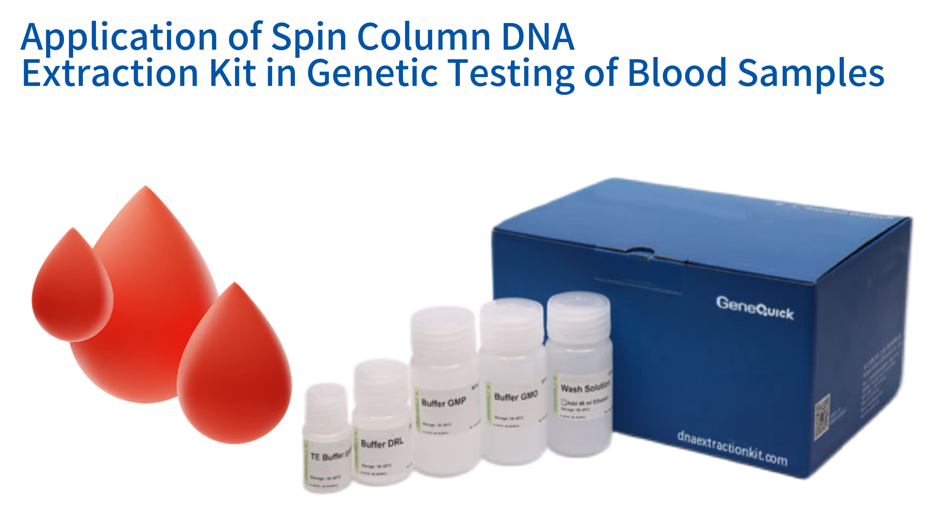
Genetic testing has become a cornerstone of modern medicine, clinical diagnostics, and life sciences research. The accuracy and reliability of these tests depend fundamentally on the quality of the starting material: DNA. Obtaining high-purity, intact DNA from blood samples, which contain a complex mixture of cells, proteins, and other inhibitors, is a critical first step. Spin column DNA extraction kits provide a standardized, efficient, and robust solution for this challenge. These kits utilize a silica membrane technology that selectively binds DNA under specific conditions, allowing for the removal of contaminants through a series of rapid centrifugation steps. The resulting DNA is free of proteins, RNA, and other molecules that could interfere with downstream applications like polymerase chain reaction (PCR) or next-generation sequencing (NGS). For clinical laboratories processing hundreds of blood samples daily, the consistency, speed, and minimal handling requirements of spin column kits make them an indispensable tool. This article provides a comprehensive exploration of how these kits work, their advantages, their application in various genetic testing scenarios, and key considerations for selection and use, ensuring that laboratories can achieve the highest standards of performance and reliability in their genetic analyses. The entire process, from a blood draw to a genetic result, hinges on this crucial extraction phase, making its mastery essential for any facility engaged in modern genetic testing.
The Fundamental Principles of Spin Column DNA Extraction Technology
Key Performance Metrics of Spin Column DNA Extraction from Blood Samples
| Performance Indicator | Typical Value | Significance for Genetic Testing |
|---|---|---|
| DNA Yield (from 200µL whole blood) | 3-8 µg | Sufficient for multiple PCR reactions or NGS library prep |
| A260/280 Ratio | 1.8-2.0 | Indicates minimal protein contamination, critical for PCR efficiency |
| A260/230 Ratio | >2.0 | Confirms removal of chaotropic salts and carbohydrates (PCR inhibitors) |
| DNA Fragment Size | 20-50 kb | High molecular weight suitable for long-range PCR and NGS |
| Processing Time per Sample | 20-40 mins | Enables high-throughput processing in clinical labs |
| PCR Inhibition Rate | <5% | Ensures reliable amplification for diagnostic testing |
Spin column technology, at its core, is a form of solid-phase extraction. It leverages the unique chemical properties of DNA and a specialized silica membrane housed within a small, disposable column. The process is elegantly simple yet highly effective, relying on controlled changes in buffer chemistry to guide DNA through four key stages: lysis, binding, washing, and elution. Understanding these stages is crucial for troubleshooting and optimizing the extraction process for specific blood sample types. The buffers in a commercial spin column dna extraction kit for blood are carefully formulated to ensure maximum DNA recovery and purity, a standard that is difficult to achieve with homemade reagents. This section details the molecular dance that occurs within the column, explaining how a complex biological fluid like blood is transformed into a pure DNA solution ready for analysis. The entire procedure typically takes less than an hour, a testament to the power of this technology.
Cell Lysis and Protein Digestion
The journey begins with the blood sample itself. Whole blood contains red blood cells, which lack nuclei and DNA, and white blood cells (leukocytes), which are the primary source of genomic DNA. The first step is to selectively lyse, or break open, the cell membranes of these leukocytes. This is achieved using a lysis buffer, which typically contains a detergent like sodium dodecyl sulfate (SDS) or Triton X-100. These detergents disrupt the lipid bilayer of the cell membrane and the nuclear envelope, releasing the cellular contents, including the DNA, into the solution. Simultaneously, the lysis buffer contains proteases, most commonly Proteinase K. This robust enzyme rapidly digests histone proteins that are tightly bound to the DNA, as well as other cellular proteins. This digestion is critical; if proteins are not removed, they can co-purify with the DNA and inhibit downstream enzymes like polymerases. The efficiency of this lysis and digestion step directly impacts the final DNA yield, with complete digestion ensuring maximum release of nucleic acids from the white blood cells.
The conditions during lysis are carefully controlled. The buffer is maintained at an alkaline pH and often includes chelating agents like EDTA. EDTA binds to magnesium ions, which are essential cofactors for DNases—enzymes that degrade DNA. By sequestering these ions, the buffer protects the released DNA from nuclease digestion, preserving its integrity. The incubation time and temperature are also critical; most protocols call for incubation at 56°C for 10-20 minutes. This elevated temperature optimizes the activity of Proteinase K and aids in denaturing proteins. For samples with a high white blood cell count, such as those from patients with certain leukemias, a longer incubation may be necessary. After this step, the lysate is a complex mixture of fragmented proteins, lipids, carbohydrates, and the liberated DNA, all suspended in the buffer. This mixture is now ready for the next phase, where DNA will be selectively captured onto the silica membrane.
DNA Binding to the Silica Membrane
The lysate is then transferred into the spin column, which contains a silica membrane. For DNA to bind to this membrane, the chemical environment must be just right. This is achieved by adding a binding buffer, which is predominantly composed of a high concentration of chaotropic salts, such as guanidine hydrochloride or guanidine thiocyanate. These salts serve two vital functions. First, they disrupt the hydrogen bonding network of water, effectively dehydrating the DNA molecules. Second, they break the hydrogen bonds between the DNA and water, encouraging the DNA to adsorb onto the silica surface. The principle behind this adsorption is that under these high-salt, dehydrating conditions, the negatively charged phosphate backbone of DNA is able to overcome its repulsion from the negatively charged silica surface. Positively charged ions in the salt solution form a bridge between the DNA and the silica, allowing the DNA to bind strongly and selectively.
The column is then placed into a collection tube and centrifuged. This forces the liquid lysate through the silica membrane. While the liquid, containing proteins, cell debris, and other contaminants, passes through into the collection tube, the DNA remains tightly bound to the membrane. The selectivity of this binding is remarkable; under these specific salt and pH conditions, RNA and other cellular components do not bind with the same affinity. This crucial separation step effectively isolates the DNA from the vast majority of biological impurities. The chaotropic salts also serve to inactivate any remaining nucleases, further protecting the DNA. The success of this binding step is determined by the quality of the membrane, the correct composition of the binding buffer, and the centrifugal force applied, ensuring that all available DNA is captured while contaminants are efficiently washed away.
Sequential Wash Steps to Remove Impurities
With the DNA securely bound to the membrane, the next phase is to wash away any lingering contaminants. This is achieved through one or two wash steps using different buffers. The first wash buffer typically contains a chaotropic salt similar to the binding buffer, but at a lower concentration, along with ethanol. This buffer is designed to remove residual proteins and carbohydrates that might have non-specifically adhered to the membrane or the DNA. As this buffer is passed through the column by centrifugation, it carries these impurities away. The ethanol in the buffer helps to keep the DNA precipitated and bound to the membrane while solubilizing other unwanted biomolecules. This step is crucial for obtaining high-purity DNA, as it strips away contaminants that could inhibit downstream enzymatic reactions like PCR. Inefficient washing can lead to DNA that appears pure by spectrophotometry but fails to amplify, a common pitfall in genetic testing.
A second wash buffer, often simply called the wash buffer, is then applied. This buffer is primarily ethanol-based, with a low salt concentration. Its function is to remove residual chaotropic salts from the membrane. Chaotropic salts are potent PCR inhibitors, so their complete removal is non-negotiable for successful genetic testing. The ethanol-based buffer efficiently washes these salts through the column. Following the addition of this buffer and a final centrifugation, the flow-through is discarded. At this point, the DNA is bound to a clean, dry membrane, free of proteins, carbohydrates, salts, and other inhibitors. However, the membrane now contains ethanol from the wash buffer. A final, dry spin (centrifugation without adding any liquid) is often performed to evaporate any residual ethanol, as alcohol can also interfere with downstream applications and prevent efficient elution of the DNA. This meticulous washing process is what ensures the final DNA product is of the highest quality.
Elution of Purified DNA
The final step is to release the purified DNA from the silica membrane. This is achieved by changing the chemical environment once again. A low-salt buffer, typically Tris-EDTA (TE) buffer or simply nuclease-free water, is applied directly onto the membrane. The absence of salt and the slightly alkaline pH (around 8.0-8.5) reverse the binding conditions. Water molecules are now able to hydrate the DNA, breaking the salt bridges that held it to the silica. The DNA rehydrates and diffuses from the membrane into the liquid buffer. During centrifugation, this DNA-rich liquid is spun out of the column and into a clean collection tube. This final eluate is the pure, ready-to-use DNA. The volume of elution buffer used is a key variable. Using a smaller volume (e.g., 50 µL) will yield a higher concentration of DNA, which is ideal for applications like NGS. Using a larger volume (e.g., 200 µL) will yield a lower concentration but may be preferred if multiple downstream assays are planned from the same sample.
The efficiency of elution can be influenced by several factors. The temperature of the elution buffer matters; pre-warming the buffer to 56°C or 70°C can significantly increase DNA yield, especially for larger fragments. Incubating the buffer on the membrane for a few minutes before centrifugation allows for more complete rehydration and release of the DNA. The integrity of the DNA at this stage is excellent. The entire extraction process, from lysis to elution, is designed to be gentle, minimizing shearing and preserving high-molecular-weight DNA. This is crucial for applications like long-range PCR or certain NGS library preparation methods. The final product, a clear, colorless solution of pure DNA in a tiny tube, is the foundation for all subsequent genetic analysis, whether it be for diagnosing a genetic disorder, identifying a pathogen, or conducting cutting-edge genomic research. The reliability of this final step is what makes the spin column format so universally adopted.
Advantages of Spin Column Kits for Blood-Derived DNA
While several methods exist for DNA extraction, the spin column format offers a unique combination of benefits that make it particularly well-suited for processing blood samples in genetic testing workflows. Its dominance in the market is a direct result of its ability to consistently deliver high-quality DNA with operational simplicity. Compared to traditional organic extraction methods like phenol-chloroform, spin columns are safer, faster, and more reproducible. They eliminate the use of hazardous organic solvents, making them ideal for clinical and routine laboratory environments. This section outlines the key advantages that solidify the spin column's role as a workhorse technology for blood-based genetic testing, from the research bench to the high-throughput diagnostic lab.
High Purity and Removal of PCR Inhibitors
Blood is a notoriously challenging sample type for PCR due to the presence of potent inhibitors. Hemoglobin and its breakdown products, such as hemin, lactoferrin, and immunoglobulins, are known to interfere with the activity of DNA polymerases. The presence of even trace amounts of these substances can lead to false-negative results or reduced amplification efficiency. The chemistry of spin column kits is specifically designed to overcome this challenge. The combination of protein digestion by Proteinase K and the selective binding of DNA to the silica membrane in the presence of chaotropic salts effectively strips away these inhibitory molecules. The subsequent wash steps, particularly those with ethanol-based buffers, ensure that any residual inhibitors are thoroughly removed before the final elution. The result is DNA with exceptional purity, typically characterized by A260/280 ratios between 1.8 and 2.0 and A260/230 ratios often exceeding 2.0, indicating minimal protein and chaotropic salt contamination, respectively. This high purity is directly correlated with robust and reliable PCR performance, a non-negotiable requirement for clinical genetic tests.
The consistency of purity across different blood samples, even those from patients with varying hematocrit levels or disease states, is another major advantage. Manual or homebrew methods often struggle with sample-to-sample variability, requiring protocol adjustments that are impractical in a busy lab. A well-formulated spin column kit, validated for use with human whole blood, delivers reliable purity batch after batch. This consistency builds confidence in the downstream results. For example, a laboratory performing genetic testing for hereditary disorders can be assured that a failed PCR reaction is likely due to the absence of the target sequence and not a problem with DNA quality. This reliability streamlines troubleshooting and reduces the need for repeat testing, saving both time and money. The proven ability of spin column kits to deliver inhibitor-free DNA is a primary reason for their widespread adoption in accredited clinical laboratories worldwide.
Speed, Efficiency, and Standardized Protocols
Time is a critical factor in many laboratory settings. For patients awaiting a genetic diagnosis, faster results can be life-changing. For research labs, quicker extraction means more experiments can be performed. Spin column kits offer a dramatic increase in speed compared to older methods. A typical protocol can be completed in 20-40 minutes, from lysed blood to purified DNA. This rapid turnaround is made possible by the centrifugal format, which quickly moves liquids through the membrane, and the use of pre-aliquoted, ready-to-use buffers. There is no need for time-consuming steps like overnight precipitation or hazardous solvent evaporation. The entire process is streamlined, allowing a single technician to process dozens of samples in a few hours. This efficiency translates directly into lower labor costs and higher laboratory throughput, enabling facilities to handle larger workloads without a proportional increase in staff.
Beyond speed, the standardized protocols are a major benefit. Each kit comes with a detailed, step-by-step instruction manual. This standardization ensures that different users, or the same user on different days, will perform the extraction in an identical manner. This is fundamental for Good Laboratory Practice (GLP) and for maintaining ISO or CAP accreditation. The protocol's simplicity also reduces the potential for operator error. With fewer pipetting steps and no hazardous chemicals, the risk of mistakes that could compromise sample integrity is minimized. This ease of use means that even technicians with minimal molecular biology experience can be quickly trained to perform high-quality DNA extractions. The combination of speed and built-in protocol standardization makes the spin column format an ideal choice for core facilities, diagnostic labs, and any setting where reproducibility and efficiency are paramount.
Scalability and Minimal Equipment Needs
Another significant advantage of spin column technology is its scalability. The basic principle is the same whether processing a single sample or 96 samples in parallel. For low-throughput needs, individual spin columns processed in a standard benchtop microcentrifuge are perfectly adequate. For higher throughput, kits are available in 96-well plate formats. These plates contain silica membranes at the bottom of each well, allowing for the simultaneous processing of 96 samples using a vacuum manifold or a centrifuge equipped with a plate rotor. This seamless scalability means that a laboratory can start with a low-throughput manual method and, as its workload grows, transition to a higher-throughput automated or semi-automated workflow without having to completely redesign its extraction process or validate a fundamentally new technology. This flexibility is invaluable for growing laboratories and core facilities.
The equipment required for spin column extraction is minimal and already present in almost every molecular biology lab. At its most basic, only a microcentrifuge, a heat block or water bath, and a set of micropipettes are needed. This low barrier to entry makes the technology accessible to labs with limited budgets, including those in educational settings or in developing regions. There is no need for expensive robotics or specialized instruments, although automation is certainly possible. This simplicity stands in stark contrast to magnetic bead-based systems, which often require specialized separation equipment, or to automated liquid handling workstations, which represent a significant capital investment. The low infrastructure requirements, combined with the high-quality output, make spin column kits the most practical and cost-effective solution for the vast majority of laboratories performing DNA extraction from blood for genetic testing. The only consumable that is regularly used up is the column itself, which is a small, disposable, and relatively inexpensive component.
Applications in Genetic Testing from Blood Samples
The purified DNA obtained from spin column extraction serves as the foundational template for an incredibly diverse range of genetic tests. From diagnosing rare inherited diseases to guiding cancer treatment, the applications are vast and growing. The quality of the DNA—its purity, integrity, and concentration—directly impacts the success and accuracy of these downstream analyses. This section explores the most common and critical applications where spin column-extracted DNA from blood is the sample of choice. The versatility of this input material underscores the importance of a reliable and high-performing extraction method. Whether the goal is to detect a single mutation or sequence an entire genome, the journey begins with a robust DNA sample, and spin columns consistently deliver.
PCR and Quantitative PCR (qPCR) for Disease Detection
Polymerase chain reaction (PCR) and its real-time counterpart, qPCR, are the most widely used techniques in molecular diagnostics. They are employed for a multitude of purposes, including detecting infectious diseases (like HIV, hepatitis B, and SARS-CoV-2), identifying genetic mutations associated with inherited disorders (such as cystic fibrosis or sickle cell anemia), and monitoring minimal residual disease in cancer patients. For all these applications, the DNA template must be free of inhibitors to ensure efficient amplification. A failed qPCR reaction due to a poor-quality sample can delay diagnosis and treatment. DNA extracted from blood using a spin column kit, particularly one optimized for clinical use, provides the clean, consistent template that these sensitive assays require. The removal of heme and other PCR inhibitors from blood is so effective that it allows for accurate quantification of viral load or gene expression levels.
The compatibility with multiplex PCR, where multiple targets are amplified in a single reaction, is another key benefit. High-quality, pure DNA ensures that all primers in the multiplex reaction can bind and extend efficiently without interference. For example, in pharmacogenomic testing, where a patient's genetic profile is used to predict their response to certain drugs, multiplex PCR panels are often used to genotype multiple relevant SNPs (single nucleotide polymorphisms) simultaneously. The success of these complex reactions hinges on the quality of the starting DNA. Using DNA from a reliable spin column kit minimizes the risk of partial or complete reaction failure, leading to more definitive and actionable results for clinicians. The robust nature of the DNA obtained makes it a versatile template for any PCR-based application.
Next-Generation Sequencing (NGS) for Comprehensive Genomic Analysis
Next-generation sequencing (NGS) has revolutionized genomic research and is rapidly being adopted into clinical practice. NGS allows for the simultaneous sequencing of millions of DNA fragments, providing a comprehensive view of a patient's genome, exome, or a targeted panel of genes. The input DNA for NGS must be of exceptional quality. It needs to be not only pure but also highly intact, as fragmentation can lead to library preparation failures and biased sequencing results. Spin column extraction is a well-established method for producing high-molecular-weight DNA suitable for NGS. The gentle handling during the binding and elution steps minimizes physical shearing, preserving long DNA fragments. This is particularly important for applications like whole-genome sequencing, where long reads are needed for accurate assembly and structural variant detection. Laboratories performing oncology research using clinical samples depend on this integrity to accurately characterize tumor genomes from blood samples.
The purity of the DNA is equally critical for NGS. Contaminants like proteins, carbohydrates, or residual chaotropic salts can interfere with the enzymatic steps of library preparation, such as end-repair, adapter ligation, and PCR amplification. These interferences can result in low library yields, adapter dimers, and poor sequencing data quality. The stringent wash steps in spin column protocols are designed to eliminate these inhibitors, ensuring that the DNA is fully compatible with NGS workflows. Furthermore, the consistency of spin column extraction is vital for large-scale sequencing projects where hundreds or thousands of samples are processed. The ability to produce uniform DNA quality across all samples reduces batch effects and increases the statistical power of the study. As NGS becomes more routine in clinical diagnostics, the demand for reliable, high-quality input DNA, such as that produced by spin column kits, will only continue to grow.
Biobanking and Long-Term Storage of Genetic Material
Biobanks are essential repositories for biological samples, supporting countless research studies aimed at understanding human health and disease. DNA extracted from blood is a core component of many biobanks. For this material to be useful for future research, often decades later, it must be stored in a way that preserves its integrity. The DNA must be stable, free from nucleases, and protected from damage. The elution buffer used in spin column kits, often TE buffer, is specifically formulated for long-term storage. The EDTA in the buffer chelates magnesium ions, inactivating any potential DNase contamination and preventing enzymatic degradation. Storing DNA at -20°C or -80°C in this buffer ensures that it remains intact and suitable for analysis for many years. The high purity achieved by spin column extraction also means that there are no residual contaminants in the stored sample that could slowly degrade the DNA over time, even at low temperatures.
The standardized format of the extracted DNA—a known concentration and volume in a standard tube—is also a significant advantage for biobanking. This uniformity simplifies sample management, tracking, and distribution. Researchers requesting samples from a biobank can be confident in the quality of the material they receive, knowing it was produced using a validated, consistent protocol. This reliability is crucial for the reproducibility of scientific research. Furthermore, many spin column kits are certified for in vitro diagnostic (IVD) use, meeting the stringent regulatory requirements for clinical biobanks. This certification ensures traceability and quality control at every step, from sample collection to DNA storage. For large-scale prospective studies that will analyze samples years after collection, the proven stability and quality of spin column-extracted DNA make it the gold standard for biobanking genetic material from blood.
Optimizing Protocols for Blood Sample Variations
While spin column kits are designed to be robust, not all blood samples are the same. Factors such as the sample volume, the type of anticoagulant used, and the quality of the blood (fresh vs. frozen) can influence extraction efficiency and DNA quality. Understanding how to optimize the protocol for these variables is key to consistently obtaining the best results. A skilled technician knows that a "one-size-fits-all" approach may not always be sufficient, especially when dealing with challenging samples. This section provides practical guidance on how to adapt the standard protocol to common variations encountered in blood samples, ensuring high yields and purity every time. These minor adjustments can make a significant difference in the success of downstream genetic tests, especially those requiring high DNA input or of exceptional quality, such as those performed on samples intended for forensic analysis.
Adjusting for Sample Volume and White Blood Cell Count
The standard protocol for a blood spin column kit is often optimized for a specific input volume, commonly 200 µL of whole blood. However, laboratories may sometimes need to process larger volumes to increase DNA yield, or smaller volumes, such as from pediatric or elderly patients where blood collection is difficult. For larger volumes, simply scaling up the lysis reagents is not always sufficient, as the silica membrane in the spin column has a finite binding capacity. Exceeding this capacity can lead to DNA breakthrough, where some DNA passes through the column without binding, resulting in a lower-than-expected yield. In such cases, it is often better to process the sample in multiple aliquots and pool the eluted DNA, or to use a kit specifically designed for larger input volumes. Conversely, for very small volumes, the main challenge is not losing the sample during the multiple pipetting and centrifugation steps. Carrier RNA is sometimes included in kits for low-input samples to improve DNA recovery by co-precipitating with the target DNA.
The white blood cell count, which can vary significantly between individuals and disease states, also impacts the process. Patients with infections or leukemias may have elevated counts, while those undergoing chemotherapy may have very low counts. For samples with very high leukocyte counts, the amount of DNA released during lysis can exceed the binding capacity of the column, even with standard input volumes. In these cases, reducing the sample volume or performing a more dilute lysis may be necessary. For samples with very low leukocyte counts (leukopenic), such as from immunocompromised patients, the DNA yield may be low. Here, maximizing recovery is key. This can involve using a larger input volume, if available, extending the Proteinase K digestion time to ensure complete lysis of all cells, and eluting the DNA in a smaller volume to increase concentration. Understanding these nuances allows the laboratorian to proactively adjust the protocol to match the sample characteristics.
Impact of Anticoagulants and Blood Storage Conditions
The type of anticoagulant used in the blood collection tube is a critical variable. The most common are EDTA, citrate, and heparin. EDTA and citrate are generally compatible with DNA extraction and downstream PCR. Heparin, however, is a potent inhibitor of PCR and should be avoided whenever possible for samples destined for genetic testing. Heparin can co-purify with DNA during extraction and will irreversibly inhibit Taq polymerase, leading to false-negative results. If heparinized blood is the only sample available, special protocols are required, often involving extensive washing or an additional heparinase treatment step, which adds time and complexity. Most spin column kits are validated for use with EDTA- or citrate-treated blood, and using these is strongly recommended. The choice of collection tube is often the first and most important pre-analytical variable a laboratory can control.
The storage and age of the blood sample before extraction is another key factor. Fresh blood, processed within a few hours of collection, will generally yield the highest quality, highest molecular weight DNA. If samples must be stored, refrigeration at 4°C for a few days is acceptable, but freezing is preferred for long-term storage. However, repeated freeze-thaw cycles are detrimental; they can cause leukocyte lysis even before the extraction begins, releasing nucleases that can degrade DNA. It is best to aliquot blood into smaller volumes before freezing if multiple tests are planned. For frozen blood, it is crucial to thaw samples slowly on ice and mix gently to avoid further shearing of DNA from lysed cells. Some protocols may recommend a longer Proteinase K digestion for previously frozen samples to ensure complete lysis and to degrade any nucleases that may have been activated. Adhering to best practices for blood storage and handling is essential to protect the integrity of the nucleic acids and ensure that the high-quality DNA obtained from the spin column is representative of the original sample.
Spin Column vs Other DNA Extraction Methods - Key Parameters Comparison
| Performance Parameter | Spin Column | Phenol-Chloroform | Magnetic Beads | Best For |
|---|---|---|---|---|
| DNA Purity (A260/280) | 1.8-2.0 | 1.7-1.9 | 1.8-2.0 | Spin Column/Magnetic Beads: Clinical diagnostics |
| Processing Time per Sample | 20-40 mins | 60-90 mins | 30-50 mins | Spin Column: Fast turnaround labs |
| Equipment Requirements | Basic | Basic | Specialized | Spin Column: Low-budget facilities |
| Hazardous Materials | None | Organic solvents | None | Spin Column/Magnetic Beads: Clinical labs |
| Automation Compatibility | Moderate | Low | High | Magnetic Beads: High-throughput labs |
| Cost per Sample | Low-Medium | Low | High | Phenol-Chloroform: Research-only labs |
Choosing the Right Spin Column Kit for Your Laboratory
With numerous commercial vendors offering spin column DNA extraction kits, selecting the right one for a specific laboratory's needs can be a daunting task. While the underlying principle is the same, kits can differ in their buffer compositions, membrane chemistry, throughput format, and intended applications. A kit that works perfectly for a research lab processing fresh animal blood may not be ideal for a clinical lab extracting DNA from archived human samples for diagnostic purposes. Making an informed choice requires a careful evaluation of several key parameters aligned with the laboratory's workflow, quality requirements, and budget. This section provides a practical framework for navigating the selection process, ensuring that the chosen kit delivers consistent, reliable performance for its intended use.
Key Performance Parameters: Yield, Purity, and Integrity
The most fundamental criteria for any extraction kit are the quality and quantity of DNA it produces. Yield refers to the amount of DNA obtained, typically measured in micrograms (µg) from a standard input volume. For most genetic tests, a minimum yield is required. For example, whole-genome sequencing may require 1-2 µg of DNA, while a standard PCR reaction may need only a few nanograms. The kit's specifications should clearly state the expected yield from a given volume of blood. Purity is assessed spectrophotometrically, with the A260/280 and A260/230 ratios serving as key indicators. A good kit should consistently produce ratios within the accepted ranges (1.8-2.0 for 260/280, >1.8 for 260/230). Any deviation suggests contamination that could affect downstream results. It is wise to look for independent validation data or published studies that use the kit, rather than relying solely on the manufacturer's claims.
DNA integrity, or the average fragment size, is another critical but sometimes overlooked parameter. For many PCR-based applications, moderately fragmented DNA is acceptable. However, for long-range PCR, optical mapping, or certain NGS library prep methods, high-molecular-weight DNA is essential. The extraction method should be gentle enough to preserve long fragments. This can be assessed by running the extracted DNA on an agarose gel; a high-quality extraction will show a single, high-molecular-weight band with minimal smearing. Kits that involve vigorous vortexing or harsh lysis conditions may lead to more shearing. For laboratories whose primary downstream application is, for example, identifying microorganisms in blood for research, moderate integrity may be sufficient. For those performing advanced genomic analyses, prioritizing a kit known for preserving DNA integrity is crucial. Evaluating all three parameters—yield, purity, and integrity—provides a complete picture of kit performance.
Throughput, Automation, and Workflow Integration
A laboratory's daily sample volume dictates the required throughput. A small research lab processing 10-20 samples a week may be perfectly served by a manual spin column kit. A large diagnostic lab processing hundreds of samples a day, however, needs a solution that can keep pace. For high-throughput environments, kits are available in 96-well plate formats that can be processed using vacuum manifolds or centrifuges. Some vendors also offer kits that are optimized for use on automated liquid handling workstations. Choosing a kit that can be automated provides a clear path for future scalability. It is important to assess not just the extraction time, but the total hands-on time required. A kit with fewer pipetting steps and ready-to-use buffers will be more efficient and less prone to error, regardless of throughput. The kit should integrate seamlessly with the laboratory's existing equipment, such as centrifuges and heat blocks.
Workflow integration also involves considering the sample types processed alongside blood. If a laboratory handles a variety of samples, such as tissue, saliva, or cultured cells, using a universal kit that can process all these sample types with minor protocol modifications can simplify inventory and training. Some manufacturers offer a "family" of kits based on the same core technology, allowing for consistency across different applications. This can be a major advantage in a multi-user core facility. Another consideration is the downstream application. If the DNA is destined for a specific NGS platform, some kit manufacturers offer validation data or even co-marketing agreements with the platform provider, guaranteeing compatibility. This level of integration can save significant time and effort in assay validation. By considering the entire workflow, from sample reception to data generation, laboratories can select a kit that not only extracts DNA well but also enhances overall operational efficiency.
Regulatory Compliance and Quality Assurance
For clinical and diagnostic laboratories, regulatory compliance is paramount. The choice of an extraction kit is not just a technical decision but a regulatory one. Kits that are labeled as "For Research Use Only" (RUO) are not validated for clinical diagnostics. For patient testing, laboratories must use kits that are certified as In Vitro Diagnostic (IVD) medical devices, bearing a CE-IVD mark in Europe or meeting FDA requirements in the United States. These kits have undergone rigorous validation to ensure their performance, safety, and consistency for clinical use. Using such kits simplifies the laboratory's own validation process and is often a requirement for accreditation. The documentation provided with IVD kits, including detailed instructions for use and certificates of analysis, supports a robust quality management system. This is a key aspect of ensuring reliable and defensible patient results. The traceability and quality control inherent in IVD-labeled products are non-negotiable in a clinical setting.
Beyond the kit's regulatory status, the manufacturer's own quality assurance practices are important. Reputable manufacturers operate under ISO 13485 standards, which mandate a comprehensive quality management system for the design and manufacture of medical devices. They will have rigorous quality control checks on every batch of kits they produce, ensuring lot-to-lot consistency. This consistency is vital for any laboratory, but especially for those performing longitudinal studies or clinical trials, where data generated months or years apart must be comparable. Asking a potential supplier for their quality control data and evidence of their manufacturing certifications is a reasonable and prudent step. For forensic laboratories, additional certifications, such as ISO 18385, which minimizes the risk of human DNA contamination in products used for forensic DNA analysis, may be required. Aligning the kit's compliance with the laboratory's own accreditation and quality standards is the final, essential step in the selection process.