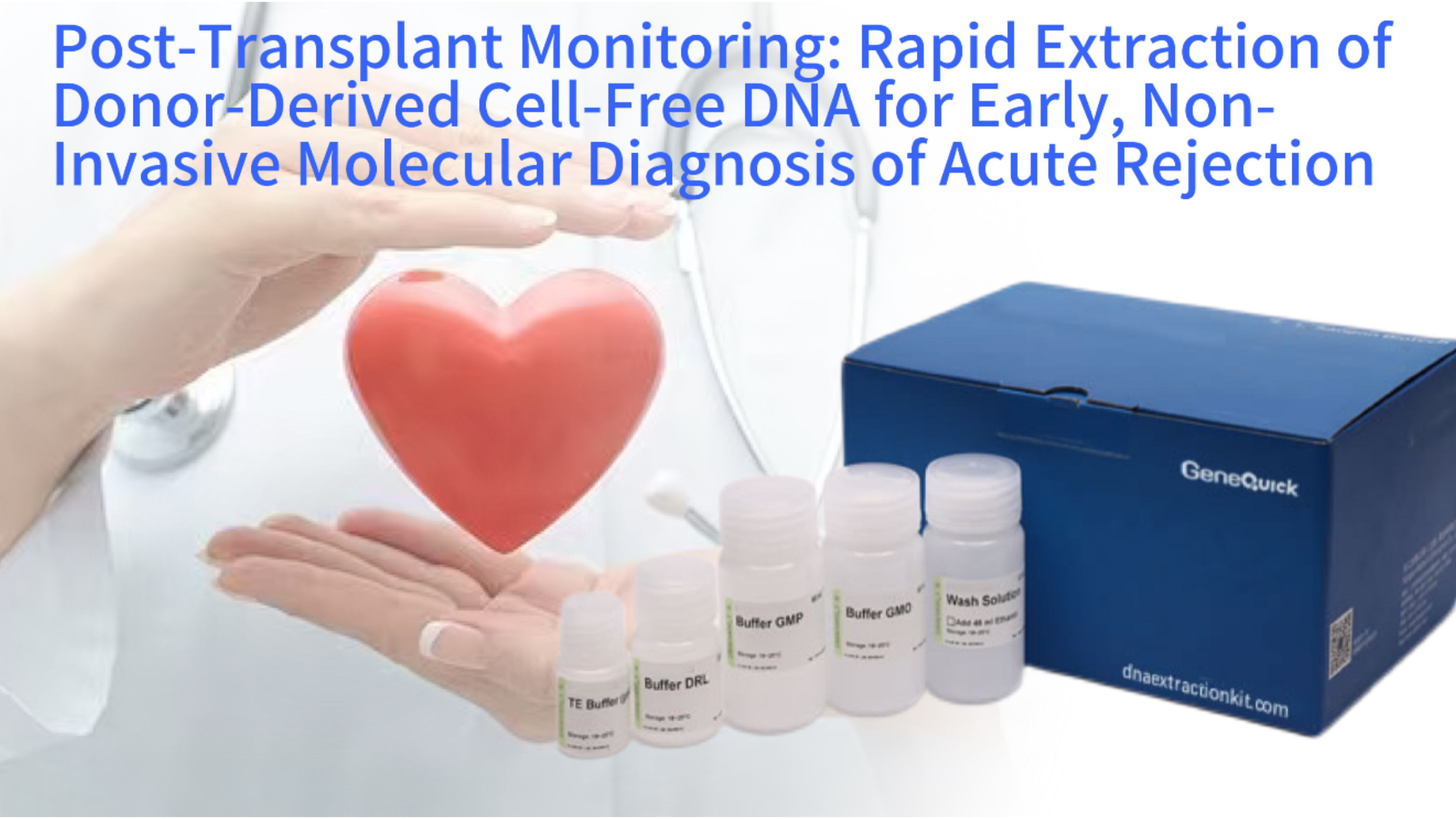
Blood Collection
Peripheral blood draw (EDTA/Streck tubes)
Plasma Isolation
Double centrifugation (cell/platelet removal)
cfDNA Extraction
Rapid magnetic bead-based purification
dd-cfDNA Quantification
ddPCR/NGS for donor fraction analysis
Clinical Interpretation
Trend analysis & graft injury assessment
Therapeutic Action
Immunosuppression adjustment if needed
Following an organ transplant, the continuous and silent battle of immune surveillance determines the long-term survival of the graft. The earliest signs of acute rejection, a process where the recipient's immune system attacks the donor organ, are subtle cellular events occurring deep within the transplanted tissue. Traditional monitoring relies on periodic biopsies, an invasive procedure with inherent risks of bleeding, infection, and sampling error that can only detect injury after significant damage has occurred. A paradigm shift is underway, moving from invasive tissue sampling to a simple blood draw. This shift is powered by the analysis of donor-derived cell-free DNA (dd-cfDNA), tiny fragments of genetic material released into the recipient's bloodstream from dying cells of the transplanted organ. The practical application of this powerful biomarker hinges on one critical laboratory step: the rapid, efficient, and highly pure extraction of cfDNA from plasma. This article explores how modern rapid DNA extraction technologies are enabling the transition of dd-cfDNA analysis from a research tool to a routine, non-invasive clinical test for the early detection of acute rejection, offering the potential to transform post-transplant patient management.
The Biological Basis: Donor-Derived Cell-Free DNA as a Liquid Biopsy for Graft Health
Cell-free DNA consists of short, fragmented DNA molecules circulating outside of cells in bodily fluids like blood plasma. In a healthy individual, cfDNA originates primarily from hematopoietic cells. Following solid organ transplantation, a second population emerges: DNA fragments from the cells of the donor organ. Under stable conditions, a low, steady-state baseline of dd-cfDNA is present, resulting from normal cellular turnover in the graft. During episodes of acute rejection, however, immune-mediated cell death causes a significant and measurable increase in the amount of dd-cfDNA released into the recipient's circulation. Studies have shown that the fraction of dd-cfDNA can rise from a baseline of less than 0.5% to over 2-5% during rejection episodes, often preceding clinical symptoms or changes in standard organ function tests by days or weeks. This makes it a highly sensitive early warning signal.
The characteristics of cfDNA present unique analytical challenges and opportunities. These molecules are typically short, with a dominant peak around 166 base pairs, corresponding to the length of DNA wrapped around a nucleosome plus linker DNA. They exist in a vast background of recipient-derived cfDNA, often at a ratio of 1:1000 or less. The half-life of cfDNA in circulation is short, approximately 15 minutes to a few hours, meaning its levels reflect real-time, dynamic processes within the graft. This rapid turnover is a key advantage for monitoring, as changes in graft status can be detected quickly, but it also imposes a requirement for stable and standardized pre-analytical sample processing to prevent rapid degradation or dilution of the signal before analysis can occur.
The Dynamics of cfDNA Release During Immune-Mediated Injury
The pathophysiology of acute rejection involves T-cell and antibody-mediated attacks on the graft's vascular endothelium and parenchymal cells. This targeted cytotoxicity leads to programmed cell death and necrosis, resulting in the release of cellular contents, including fragmented genomic DNA, into the interstitial fluid and subsequently into the bloodstream. The quantity of dd-cfDNA correlates with the severity and extent of the injury. Research published in journals like *Transplantation* indicates that not only does the total quantity increase, but the size profile of the dd-cfDNA may also shift during rejection, potentially offering an additional layer of diagnostic information. The release is a continuous process during active injury, making serial monitoring, perhaps weekly or bi-weekly in high-risk patients, a powerful strategy for tracking graft health over time.
Advantages Over Traditional Invasive Biopsy
The percutaneous needle biopsy, the long-standing gold standard, has several limitations. It is invasive, carrying risks that patients must undergo repeatedly. It samples only a tiny, approximately 1-2 cubic centimeter, portion of a large organ, leading to potential sampling error and missing focal rejection. Histological interpretation can have variability between pathologists. Most importantly, biopsy detects injury only after it has become histologically apparent. In contrast, a liquid biopsy based on dd-cfDNA is minimally invasive, requiring only a peripheral blood draw. It interrogates the entire organ, as cfDNA is released from all injured tissues into the systemic circulation. It provides a quantitative, objective measurement and has the potential to detect immune activity at a molecular level before irreversible tissue architecture damage occurs, enabling pre-emptive therapeutic intervention.
Technical Imperative: The Need for Rapid and Specialized cfDNA Extraction
The journey from a blood sample to a dd-cfDNA fraction result is a chain of highly specialized procedures. The first and most foundational link is the extraction of total cfDNA from plasma. This is not a routine genomic DNA extraction. The target molecules are ultra-short, low-abundance, and swim in a sea of proteins, lipids, and potential PCR inhibitors like hemoglobin and immunoglobulin G. The extraction method must be exquisitely optimized for short fragments to avoid size bias that could skew the final dd-cfDNA percentage. Furthermore, the workflow from blood draw to frozen plasma must be swift and standardized to prevent the release of genomic DNA from lysing blood cells, which would massively dilute the dd-cfDNA signal with recipient background noise.
Speed in the extraction process itself is not merely a convenience; it is a component of assay robustness and clinical utility. In a busy clinical laboratory handling dozens of samples, a protocol that takes 4 hours is less feasible than one taking 45 minutes. Rapid extraction minimizes hands-on time, reduces the window for procedural errors, and enables higher throughput. More critically, many rapid extraction kits are designed for seamless integration into automated liquid handling platforms. Automation ensures exceptional reproducibility, a non-negotiable requirement for a quantitative diagnostic test where a change from 0.3% to 0.5% dd-cfDNA may be clinically significant. The choice of extraction technology directly impacts the sensitivity, specificity, and precision of the entire monitoring assay.
Key Performance Metrics of cfDNA Extraction Technologies
Challenges of Isulating Low-Abundance, Short-Fragment DNA
Conventional DNA extraction methods, often designed for long, high-molecular-weight genomic DNA from cells, can be inefficient for cfDNA. Silica-column methods may lose a significant proportion of fragments under 100 base pairs during the binding and washing steps. The binding chemistry must be tuned to capture these short molecules with high efficiency. Additionally, the elution volume must be small to concentrate the scant amount of cfDNA into a detectable range for downstream quantification, which is typically done via sensitive digital PCR or next-generation sequencing. Any inefficiency or bias in extraction directly translates into a loss of sensitivity for detecting early, low-level rises in dd-cfDNA, potentially missing the window for earliest intervention.
Integration into High-Throughput Clinical Laboratory Workflows
For a test to be adopted widely in transplant centers, it must fit into the operational flow of a clinical laboratory. This means batch processing, compatibility with common sample tube types (like Streck or EDTA tubes for plasma collection), and a turnaround time that supports clinical decision-making. Rapid extraction kits formatted for 96-well plates are ideal. They allow a technician to process 96 patient samples in roughly the same time it might take to manually process 10 using older methods. The pre-aliquoted, stable reagents in these kits reduce preparation time and minimize variability. This high-throughput, standardized approach is what makes frequent, routine monitoring of a large patient population logistically and economically viable, moving from a niche test to a standard of care.
The Core Technology: Principles of Rapid cfDNA Extraction from Plasma
Modern rapid extraction kits for cfDNA overwhelmingly utilize silica-based magnetic bead technology. The process begins with plasma, the cell-free fraction of blood obtained by a double centrifugation protocol to ensure all cells and platelets are removed. The plasma is mixed with a lysis-binding buffer containing chaotropic salts, such as guanidine hydrochloride, and a detergent. This buffer serves a dual purpose: it denatures and dissolves any residual protein complexes that might be shielding cfDNA, and it creates the high-ionic-strength, low-pH environment necessary for DNA adsorption to silica. Importantly, no mechanical disruption is needed, as the DNA is already free in solution.
Silica-coated magnetic beads are then added to the lysate. The chaotropic environment dehydrates the DNA molecules and the bead surfaces, promoting hydrogen bonding between the DNA phosphate backbone and the silica's hydroxyl groups. This binding is largely length-independent when optimized, capturing both long and short fragments efficiently. A magnet is applied to the tube or plate, pulling the bead-DNA complexes to the side and allowing the contaminated supernatant, now containing proteins, lipids, and other small molecules, to be completely removed. Subsequent wash steps with an ethanol-based buffer remove salts and residual impurities. Finally, a low-ionic-strength elution buffer is added. This buffer rehydrates the DNA, disrupting the hydrogen bonds and releasing pure, short-fragment-enriched cfDNA into solution, ready for precise quantification and analysis.
Magnetic Bead cfDNA Extraction Process
Lysis & Binding
Chaotropic buffer + magnetic beads
Magnetic Separation
Bead-DNA pellet formation
Washing
Ethanol-based buffer cleanup
Elution
Low-salt buffer releases cfDNA
Optimized for short-fragment (50-200bp) cfDNA recovery with minimal inhibitor carryover
Optimized Binding Chemistry for Short Fragments
The key to efficient short-fragment recovery lies in the surface chemistry of the beads and the composition of the binding buffer. Some advanced kits use beads with a carefully controlled pore size or a specialized surface coating that increases accessibility and binding affinity for smaller DNA molecules. The concentration of chaotropic salts and the pH are fine-tuned to maximize the interaction kinetics for fragments as short as 50-100 base pairs. Protocols often include a defined incubation period with gentle mixing to ensure equilibrium binding, giving even the smallest fragments ample opportunity to encounter and adhere to a bead. This optimization is empirically derived and represents a significant portion of the proprietary value of a high-performance rapid cfDNA extraction kit.
Automation-Friendly Magnetic Bead Separation
The magnetic bead platform is the enabler of speed and automation. Unlike spin columns that require centrifugation and multiple manual tube-to-tube transfers, magnetic separation is performed in a single well or tube. Liquid handling robots equipped with magnetic modules can process entire 96-well plates by sequentially adding reagents, mixing, engaging magnets to capture beads, and aspirating waste supernatants. This closed-tube workflow minimizes aerosol generation, a critical factor for preventing cross-contamination between samples when measuring very low dd-cfDNA fractions. The reproducibility of automated pipetting and timing far exceeds manual processing, directly improving the inter-assay precision of the final dd-cfDNA measurement, which is essential for tracking minute changes in a patient over time.
From Extraction to Diagnosis: Quantifying Donor-Derived Fraction
Once purified, the cfDNA must be analyzed to determine the proportion originating from the donor. Two primary technologies are used, both requiring high-quality, inhibitor-free template from the extraction step. The first is quantitative PCR-based methods, notably droplet digital PCR (ddPCR). This technique partitions a single PCR reaction into tens of thousands of nanodroplets, effectively creating individual micro-reactions. By using TaqMan probes specific for single nucleotide polymorphisms or short insertion/deletion markers that differ between donor and recipient, the absolute number of donor and recipient DNA molecules can be counted. The dd-cfDNA fraction is then calculated as [donor copies / (donor copies + recipient copies)] x 100%. ddPCR is highly precise and sensitive, capable of reliably detecting fractions below 0.1%.
The second, more comprehensive approach is next-generation sequencing (NGS). After extraction, cfDNA is converted into a sequencing library. Through shallow whole-genome sequencing or targeted sequencing of known polymorphic sites, millions of DNA fragments are mapped back to the genome. Bioinformatics algorithms then compare the alleles at these sites to a genotyping reference from the donor and recipient. The statistical power of sequencing hundreds to thousands of sites allows for extremely precise fraction calculation, often with a lower limit of detection around 0.2%. NGS can also provide additional information, such as genome-wide copy number variations that might indicate a different type of graft injury. The success of both ddPCR and NGS is utterly dependent on the initial extraction yielding cfDNA free of substances that inhibit polymerase or ligase enzymes.
ddPCR vs NGS for dd-cfDNA Quantification
Droplet Digital PCR for Absolute and Sensitive Quantification
DdPCR's strength in this application is its resistance to PCR efficiency variations and its ability to provide an absolute count without a standard curve. For transplant monitoring, assays are designed around informative SNPs. A recipient who is homozygous for allele A (AA) receiving an organ from a donor homozygous for allele B (BB) provides a simple model. Probes specific for A and B allow each droplet to be classified as recipient-positive, donor-positive, or both. The ratio of donor-positive droplets to total positive droplets gives the fraction directly. This method is robust, relatively fast, and ideal for monitoring a known donor-recipient pair over time. The cleanliness of the extracted DNA is paramount, as any inhibitor that reduces PCR efficiency could cause underestimation of one population, skewing the fraction calculation.
Next-Generation Sequencing for a Comprehensive Genomic View
NGS-based approaches offer a broader view. Targeted panels sequencing several hundred known polymorphic loci can achieve high precision. Alternatively, shallow whole-genome sequencing at low coverage analyzes millions of random fragments across the genome, identifying donor-specific alleles based on statistical overrepresentation. A major advantage of NGS is that it does not require prior knowledge of donor-specific SNPs; it can infer them from the sequencing data itself by identifying alleles in the cfDNA that are not present in the recipient's germline genotype. This is particularly useful in living donor situations where pre-transplant genotyping of the donor may not be available. The extracted cfDNA must be of sufficient quality and quantity for library preparation, which involves end-repair, adapter ligation, and amplification steps, all of which are sensitive to impurities.
Implementing a Clinical Monitoring Program: Practical Considerations
Establishing a dd-cfDNA monitoring service requires careful attention to the entire pre-analytical and analytical pipeline. It starts with phlebotomy and sample transport. Blood must be drawn into specialized tubes that stabilize nucleated blood cells, preventing them from lysing during transport and release genomic DNA. Tubes containing cell-stabilizing reagents are the standard. The time from draw to plasma processing must be tightly controlled, typically within a pre-validated window of a few days. Plasma separation via a standardized double-spin centrifugation protocol is critical to achieve platelet-poor plasma, as platelets contain genomic DNA that can lyse and contaminate the cfDNA pool.
The extraction and analysis must operate under a clinical laboratory quality management system, often seeking accreditation under standards like ISO 15189 or CAP. This involves rigorous validation of the entire assay, establishing performance characteristics such as precision, accuracy, reportable range, and reference intervals. A key part of the validation is determining the biological variation and baseline in stable transplant patients to define a "normal" range. Clinical decision thresholds for triggering concern must be established, often through longitudinal clinical outcome studies. For example, a value persistently above 1% dd-cfDNA might prompt further investigation, even in the absence of symptoms. The rapid extraction kit chosen must have demonstrated lot-to-lot consistency and be supported by the manufacturer with detailed performance data suitable for clinical validation.
Clinical dd-cfDNA Monitoring Protocol
Establish baseline dd-cfDNA (0.2-0.5%) in stable post-transplant period (4-6 weeks)
Serial monitoring (weekly/monthly) with rapid cfDNA extraction & quantification
Alert threshold: ≥2x baseline increase OR absolute value >1% dd-cfDNA
Targeted clinical investigation (biopsy/imaging) for positive alerts
Adjust immunosuppression based on combined clinical/molecular findings
Standardization of Pre-Analytical Blood Collection and Processing
The pre-analytical phase is the largest source of potential error. Protocols must specify the exact tube type, fill volume, gentle inversion steps, maximum transport temperature and time, and centrifugation speed and duration. Deviations can lead to hemolysis, which releases hemoglobin and genomic DNA, or incomplete platelet removal, both of which can ruin the test. Laboratories often create detailed manuals and train client phlebotomy sites to ensure compliance. Implementing a tracking system that logs all these variables for each sample is essential for troubleshooting and maintaining quality. The investment in standardizing this front-end process ensures that the sophisticated downstream extraction and analysis are performed on a consistent and reliable starting material.
Establishing Clinical Thresholds and Interpreting Serial Results
The interpretation of dd-cfDNA levels is dynamic and patient-specific. An absolute threshold is less informative than a patient's individual trend. A baseline is established from one or more samples drawn during a period of clinical quiescence, confirmed by stable organ function and often a protocol biopsy showing no rejection. Subsequent results are compared to this personal baseline. A sudden two-fold or greater increase, or a sustained elevation above a population-derived threshold, is considered suspicious for graft injury. It is crucial to understand that dd-cfDNA is a marker of cellular injury, not exclusively immune rejection. Other causes like infection, ischemia-reperfusion injury, or drug toxicity can also elevate levels. Therefore, a positive dd-cfDNA test is a sensitive indicator that something is wrong with the graft, prompting further investigation which may include a biopsy to determine the exact cause, but now performed in a targeted, informed manner rather than a blind screening procedure.
Future Directions and Integration with Transplant Management
The field of dd-cfDNA monitoring is rapidly evolving. Current research is focused on increasing the granularity of information obtained. One direction is methylation profiling of the extracted cfDNA. The methylation patterns are tissue-specific. By analyzing these patterns, it may become possible to not only detect that graft injury is occurring but to infer the specific cell type of origin, potentially distinguishing between rejection affecting kidney tubules versus the vascular endothelium, for example. This would provide even more precise diagnostic information to guide therapy. Another frontier is the analysis of other analytes in the same plasma sample, such as donor-derived cell-free RNA or proteins, to build a multi-omic signature of rejection.
Ultimately, the goal is to integrate dd-cfDNA monitoring into a comprehensive, personalized management algorithm for transplant recipients. Frequent, non-invasive testing could allow for immunosuppressive drug regimens to be tailored more dynamically. A rising dd-cfDNA level might prompt a pre-emptive increase in immunosuppression to avert full-blown rejection, while a persistently low level might allow for safe dose reduction to minimize drug side effects like infection, diabetes, or nephrotoxicity. This approach, known as biomarker-guided immunosuppression, promises to improve long-term graft survival and patient quality of life. The enabling technology at the heart of this personalized future remains the reliable, rapid, and high-throughput extraction of pure cfDNA, turning a simple blood sample into a rich window onto the health of the transplanted organ.
Multi-Omic cfDNA/cfRNA Analysis
1. cfDNA Quantification
Donor fraction (dd-cfDNA %) - Cell death marker
2. Epigenetic Profiling
Methylation signatures - Tissue-specific injury
3. cfRNA Expression
Immune activation pathways - Rejection mechanism
4. Integrated Signature
Specificity for rejection vs infection/toxicity
Multi-Analyte and Epigenetic Analysis for Enhanced Specificity
Beyond simple quantification, the future lies in characterization. Advanced bisulfite sequencing of extracted cfDNA can reveal its tissue of origin based on methylation signatures. A liver has a different methylation "footprint" than blood cells. Sophisticated bioinformatics can deconvolute a mixture of cfDNA fragments to estimate the percentage coming from the liver graft versus the recipient's blood cells. This approach, still largely in research, could specifically identify graft-derived molecules, potentially offering even greater specificity for rejection over other causes of injury. Similarly, extracting and analyzing cfRNA from the same plasma sample could provide information about active gene expression pathways within the graft, moving from a marker of cell death to a marker of immune activation.
Toward Personalized, Biomarker-Guided Immunosuppression Protocols
The true transformative potential of dd-cfDNA monitoring is therapeutic. Current immunosuppression is often based on population-based protocols with fixed drug levels, leading to over-treatment in some and under-treatment in others. A management strategy using dd-cfDNA as a dynamic guide is being investigated in clinical trials. In this model, a patient's immunosuppression is adjusted not just based on drug trough levels or creatinine, but on the direct molecular signal from the graft. A low or stable dd-cfDNA might support a reduction in drug dosage to alleviate side effects. A rising trend, even within the "normal" range for the population, could trigger closer monitoring or a protocol-specified intervention in a specific patient. This shift from reactive to proactive, pre-emptive care, powered by reliable molecular diagnostics, represents the next chapter in precision medicine for transplantation.