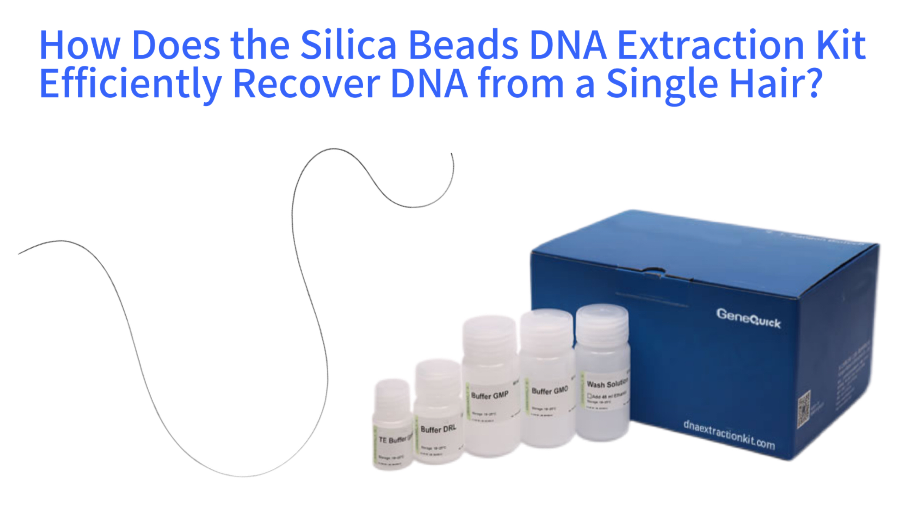
Sample Preparation
Clean & section the hair to expose DNA-rich areas
Lysis
Digest keratin with proteinase K & chaotropic salts
DNA Binding
Silica beads capture DNA in high-salt environment
Washing
Remove inhibitors (melanin, keratin fragments)
Elution
Release pure DNA into low-salt buffer
Analysis
PCR amplification & genetic profiling
Recovering DNA from a single hair represents one of the most demanding tasks in genetic analysis. Unlike a blood sample rich in nucleated cells, a hair shaft, particularly the telogen hair most commonly shed, contains a minuscule amount of nuclear DNA trapped within a resilient keratin structure. Success hinges not just on brute-force extraction but on a method that maximizes the binding and recovery of every available DNA molecule while aggressively removing inhibitors. The silica beads DNA extraction kit has emerged as the method of choice for this challenge. Its efficiency stems from a fundamental synergy between the physical properties of silica beads and the biochemical optimization of reagents designed for trace and degraded samples. This article delves into the molecular journey of DNA from a hair follicle and shaft into a purified elution tube. We will explore the unique challenges posed by hair as a biological sample, the precise chemistry that allows silica to capture DNA, the optimized step-by-step protocol for single-hair processing, and the stringent quality controls necessary for downstream forensic or diagnostic applications.
The Unique Challenge of the Single Hair as a DNA Source
A single hair is a forensically and genetically valuable sample precisely because it is often shed without notice, but its biochemical composition presents a series of extraction hurdles. The primary source of nuclear DNA resides in the follicular tag, the translucent bulb of cellular material at the root. In anagen hairs, this bulb is rich in cells. However, the more commonly found telogen hairs have a small, keratinized bulb with far fewer intact cells. When the root is absent, analysis must rely on the hair shaft, where trace amounts of nuclear DNA and a higher proportion of mitochondrial DNA are embedded within the keratinized cortex. This DNA is not only scarce but also often degraded and tightly bound within a dense protein matrix.
The challenge is therefore two-fold: accessing the DNA and purifying it efficiently from an overwhelming background of co-extracted materials. The hair's keratin structure is highly resistant to standard lysis buffers. Furthermore, during extraction, substances like melanin and other hair shaft proteins can be co-purified. These act as potent inhibitors for the polymerases used in genetic testing methods like PCR, potentially leading to complete reaction failure. The goal of any extraction protocol is to achieve total cellular and keratin breakdown to liberate DNA, followed by a purification process so selective that it captures the nanograms of DNA while leaving micrograms of keratin and inhibitors behind. The silica beads method is engineered specifically for this high-selectivity, high-recovery purification.
Anatomical Sources of DNA in a Hair Strand
The quantity and quality of DNA recoverable from a hair are entirely dependent on its anatomical segment. The follicular tag, if present, is the gold standard target. It contains the highest concentration of intact nucleated cells. When only the shaft is available, nuclear DNA is limited to the residual nuclear material from the keratinization process, often yielding less than 100 picograms of DNA. In contrast, mitochondrial DNA, present in hundreds to thousands of copies per cell, is more abundant in the shaft. This makes mitochondrial DNA analysis a viable, albeit more complex, pathway when nuclear DNA recovery fails. A robust extraction method must be capable of lysing the tough cuticle and cortex of the shaft to access this internal reservoir without destroying the fragile DNA within.
Primary Contaminants and PCR Inhibitors in Hair Samples
Keratin is the dominant protein, but it is not the only complicating factor. The hair shaft contains structural lipids and pigments, most notably melanin. Melanin is particularly problematic because it chemically inhibits the polymerase chain reaction by binding to the enzyme itself. Other potential contaminants include hair care products, environmental dirt, and bacterial DNA from the scalp microbiome. An inefficient extraction that co-purifies these substances will result in a DNA solution that may spectrophotometrically appear pure but is functionally useless for amplification. The washing stages in a silica-based protocol are therefore not mere cleaning steps; they are critical, selective barriers designed to solubilize and wash away these specific inhibitor classes while the DNA remains firmly bound to the solid silica phase.
The Molecular Mechanism: Silica-DNA Binding Chemistry
The core innovation of the silica-based method lies in its exploitation of a specific physicochemical interaction under controlled buffer conditions. This process transcends simple filtration. It is a reversible binding event driven by chaotropic salts, most commonly guanidinium thiocyanate or hydrochloride. In the context of a single hair extraction, the lysis buffer performs the dual function of denaturing the tough keratin proteins and creating the high-ionic-strength, low-pH environment necessary for the binding reaction. As the hair structure dissolves, the released DNA molecules become dehydrated and adopt a condensed conformation.
In this state, the negatively charged phosphate backbone of the DNA is shielded by the chaotropic ions. The silica bead surface, studded with hydroxyl groups, facilitates hydrogen bonding with the DNA molecule. Essentially, the chaotropic salt chaotically disrupts the hydration shells around both the silica and the DNA, allowing them to form intimate contact and bind. This interaction is highly selective in the proper buffer. Proteins, which lack a uniform, repetitive charged structure like the DNA backbone, do not bind as efficiently. Many smaller organic inhibitor molecules remain in solution. This selectivity is the first major purification event, occurring the moment the DNA contacts the bead surface amidst the chaotic lysate.
Silica-DNA Binding Mechanism
Hydrated State
DNA & silica beads surrounded by water molecules
Chaotropic Salt Addition
Disrupts water hydration layers
DNA Binding
DNA forms hydrogen bonds with silica surface
Guanidinium salts neutralize DNA charge & remove hydration layers, enabling selective binding to silica beads
The Role of Chaotropic Salts in Lysis and Binding
Chaotropic salts like guanidinium are the workhorses of the process. Their action is multifactorial. Firstly, they are potent protein denaturants, unraveling the keratin's strong tertiary structure. Secondly, they inactivate nucleases that would otherwise degrade the precious DNA upon its release. Thirdly, and most critically for the silica method, they enable the binding itself. Guanidinium ions disrupt the organized network of water molecules surrounding both the silica beads and the DNA. By removing this hydration layer, they reduce the energetic barrier for the DNA to approach the silica surface and form multiple hydrogen bonds. The high salt concentration also neutralizes the electrostatic repulsion that would normally occur between the negatively charged DNA and the slightly negative silica surface at neutral pH.
Surface Chemistry of Silica Beads for Maximized DNA Adsorption
The physical form of the silica is as important as the chemistry. Unlike the flat, two-dimensional surface of a spin column membrane, porous silica beads provide a massive three-dimensional binding surface area. A single milliliter of bead suspension can have a total surface area exceeding several square meters. This vast area dramatically increases the probability that a single, fragmented DNA molecule from a degraded hair shaft will encounter and bind to an available site. The bead size and pore structure are engineered to optimize this interaction, balancing fast diffusion of DNA into the pores with strong retention during the subsequent vigorous washing steps. This high binding capacity is essential for capturing the totality of DNA from a limiting sample where every molecule counts.
Optimized Protocol for Single-Hair DNA Extraction
The theoretical efficiency of silica binding must be translated into a meticulous practical protocol to succeed with a single hair. The process begins not with the beads, but with sample preparation. The hair is typically cleaned to remove surface contaminants, then sectioned. Cutting the hair into small segments, especially focusing on the root if present, dramatically increases the surface area exposed to the lysis buffer. The hair is then transferred to a microtube containing a specialized, highly concentrated lysis buffer. This buffer contains not only chaotropic salts but also a strong detergent and often proteinase K, an enzyme that digests keratin and other proteins into smaller peptides. An extended incubation period at 56°C with agitation is non-negotiable to ensure complete dissolution of the hair structure.
Only after a homogeneous lysate is achieved are the silica beads introduced. The binding incubation allows time for the DNA to diffuse and adhere to the bead surfaces. The critical purification phase follows: a series of washes. These are not gentle rinses. Using wash buffers containing ethanol and specific salts, the bead-DNA complex is vigorously resuspended and then immobilized, often using a magnet for magnetic silica beads. The supernatant, now containing dissolved keratin fragments, residual pigments, salts, and other small molecules, is meticulously removed. Each wash is a trade-off, designed to remove a specific class of contaminant without stripping the bound DNA. The final elution uses a low-ionic-strength buffer or nuclease-free water. This reversal of the binding conditions rehydrates the DNA, disrupts the hydrogen bonds, and releases the purified DNA into a clean, inhibitor-free solution ready for analysis.
Optimized Single-Hair DNA Extraction Protocol
Pre-Lysis Preparation
- Clean hair with sterile water/detergent
- Cut into 2-3mm segments
- Focus on root
area if present
Lysis Incubation
- Add 100μL lysis buffer + proteinase K
- Incubate at 56°C overnight with agitation
-
Ensure complete hair dissolution
DNA Binding
- Add 20μL silica beads
- Incubate at room temp for 10min
- Mix every 2min to ensure
binding
Washing Steps (3x)
- Apply magnet to pellet beads
- Remove supernatant
- Add 150μL wash buffer
- Resuspend
& repeat
Elution
- Add 10-25μL pre-warmed elution buffer
- Incubate at 65°C for 5min
- Collect eluate for
analysis
Critical Pre-Lysis Steps: Cleaning and Sectioning the Hair
Pre-analytical handling directly dictates extraction yield. A hair recovered from a scene may carry dust, oils, or other environmental inhibitors. A brief wash in a mild detergent or sterile water is common. Sectioning the hair, particularly the root bulb, with sterile scalpels or scissors is perhaps the most impactful manual step. It breaches the protective cuticle layer, allowing the lysis buffer immediate access to the internal cortical cells and DNA. For forensic hair samples where the root is absent, finely cutting the entire shaft into millimeter-long fragments is essential to maximize exposure of the internal keratin-DNA matrix to the digestive enzymes and chaotropic agents.
Adapting Incubation Times and Temperatures for Complete Keratin Breakdown
Standard tissue lysis protocols may last 30 minutes to an hour. For a single hair, especially a thick or pigmented one, this is often insufficient. Overnight digestion with proteinase K at 56°C is a routine and recommended adaptation when dealing with a single, precious hair sample. The goal is visual dissolution. The temperature is kept at 56°C to maintain optimal proteinase K activity while minimizing thermal degradation of the already vulnerable DNA. Agitation, either through constant shaking or periodic vortexing, ensures the hair segments remain immersed and the digestion enzymes circulate. Patience at this stage is the key to liberating the maximum amount of DNA for the subsequent binding step.
Maximizing Yield and Purity from a Trace Sample
When the starting material is a single hair, every technical decision must be geared toward minimizing loss and preserving the integrity of the DNA. The choice of silica bead format is pivotal. Magnetic silica beads have become the standard for high-efficiency trace extraction. Their primary advantage is the elimination of centrifugation and liquid transfer steps associated with spin columns, which can lead to bead or sample loss. With magnetic beads, the entire process from binding to elution can be performed in a single tube by simply applying and removing a magnet. This "closed-tube" approach minimizes handling, reduces the risk of contamination, and most importantly, maximizes the recovery of the bead-bound DNA complex.
Further optimizations focus on the elution step. Since the total DNA mass is low, the elution volume must be small to achieve a concentration suitable for analysis, often 10 to 25 microliters. Pre-warming the elution buffer to 55-70°C can significantly increase the efficiency of DNA release from the beads. Some protocols recommend a two-stage elution, where a first small volume of buffer is used to displace the most loosely bound DNA, followed by a brief incubation and a second elution with fresh buffer to recover the remainder. After elution, the DNA is often concentrated and further purified using specialized clean-up kits designed for forensic or ancient DNA to remove any final traces of inhibitors, ensuring the sample is pristine for sensitive downstream applications like capillary electrophoresis for STR profiling.
Advantages of Magnetic Silica Beads for Low-Volume Handling
The magnetic separation platform is transformative for trace evidence. After binding, a magnet is placed against the side of the tube, pulling the beads to the wall and forming a pellet within seconds. The contaminated supernatant can be pipetted away without disturbing the pellet. For washing, fresh buffer is added, the magnet is removed to resuspend the beads, and then reapplied to pellet them again. This process eliminates the need for spinning columns, filtering, or decanting, operations where invisible pellets can be easily lost. The final elution is performed directly onto the bead pellet in the original tube, ensuring that all DNA released from the beads is collected. This method routinely achieves recovery rates above 80% of available DNA, a critical figure when working at the limits of detection.
Strategies for Final Elution and DNA Concentration
The elution buffer is deliberately simple—typically TE buffer or water. Its low ionic strength and slightly alkaline pH (around 8.0) disrupt the hydrogen-bonding network. Heat assists this process by increasing molecular motion. Allowing the beads to incubate in the warm elution buffer for five to ten minutes, rather than immediate pelleting, gives the DNA time to diffuse away into solution. For the most challenging samples, the eluate from the first extraction can be passed over a fresh, small bed of silica beads in a second binding step. This "double purification" can further concentrate the DNA and remove any lingering inhibitors that may have carried over, though it risks some loss. The choice between a single, careful elution and a more complex concentration protocol depends on the specific sensitivity requirements of the downstream analysis.
Validation and Application in Forensic and Genetic Contexts
The ultimate test of a single-hair DNA extraction is its performance in real-world analytical pipelines. In forensic science, the extracted DNA must be of sufficient quality and quantity to generate a complete Short Tandem Repeat profile, the standard for human identification. Laboratories validate their silica bead extraction kits for hair samples according to international standards, such as the FBI's Quality Assurance Standards or ISO 18385, which governs forensic products to minimize contamination risk. This validation involves testing known samples, establishing minimum detection thresholds, and demonstrating that the method consistently produces inhibitor-free DNA that performs reliably in PCR amplification.
Beyond human identification, the same principles apply to wildlife forensics using shed fur, to archaeological studies of ancient hair, and to diagnostic scenarios where hair may be a non-invasive source of DNA for genetic disorder testing. In each case, the success of downstream techniques—whether Sanger sequencing, next-generation sequencing for mitochondrial DNA analysis, or mutation analysis—is entirely dependent on the initial extraction's success. A silica beads extraction that yields DNA with an A260/A280 purity ratio between 1.7 and 2.0 and shows no signs of inhibition in a quantitative PCR assay is considered a robust foundation for any of these advanced applications. The method's scalability also allows it to be automated on liquid handling platforms, making it suitable for processing multiple hairs from a single case or from large-scale studies efficiently and reproducibly.
Forensic Validation and STR Analysis Workflow
1. Forensic Validation
Contamination Control
Minimum DNA Input
PCR Compatibility
Consistent Results
2. STR Analysis
PCR Amplification
Capillary Electrophoresis
STR Profile Analysis
Meeting Stringent Forensic Quality Assurance Standards
Forensic applications demand more than just technical success; they require documented, auditable processes that ensure result integrity. Kits used in this field often carry certifications like ISO 18385 compliance, meaning their manufacturing process is controlled to reduce human DNA contamination. The validation study for a single-hair protocol will include sensitivity studies to define the minimum hair fragment size for reliable profiling, stochastic studies to ensure consistent results from low-template DNA, and inhibition studies using common environmental contaminants. The protocol itself must be written to eliminate cross-contamination, mandating the use of sterile, single-use consumables, dedicated workspaces, and appropriate negative controls processed alongside every batch of evidence samples.
Downstream Success: PCR Amplification and STR Profiling
The final measure of extraction efficiency is a successful electropherogram. Modern forensic PCR kits are exquisitely sensitive, capable of amplifying partial profiles from just a few dozen picograms of DNA. The role of the silica bead extraction is to deliver that DNA free of co-purified debris that would block the polymerase. Indicators of a high-quality extraction include balanced peak heights in the STR profile, the absence of elevated baseline noise, and a clean result from the internal PCR control. When an extraction from a single, rootless hair shaft produces a full, single-source DNA profile, it demonstrates the complete efficacy of the method—from breaking down the resilient keratin, capturing the fragmented DNA, removing melanin and other inhibitors, and delivering amplifiable nucleic acid. This end result is the practical proof of the underlying chemistry and optimized protocol working in concert.
Comparative Advantages and Future Directions
The silica beads method does not exist in a vacuum. It is one of several core technologies for DNA extraction. Compared to traditional organic extraction using phenol-chloroform, the silica method is far safer, faster, and more amenable to automation, with no hazardous waste. Compared to spin-column kits, which also use silica membranes, the bead format offers superior binding capacity and more efficient washing due to the three-dimensional interaction in a liquid suspension. While spin columns are excellent for many applications, the bead-based system often demonstrates a higher recovery rate from difficult, matrix-rich samples like hair, soil, or bone.
Looking forward, the field continues to evolve. Next-generation silica bead formulations are exploring surface modifications with specific functional groups to enhance binding selectivity or to preferentially bind longer DNA fragments. The integration of the method into microfluidic "lab-on-a-chip" devices promises to further minimize sample handling and loss, potentially allowing DNA extraction and analysis from a single hair fragment in a fully automated, closed system. Furthermore, the rise of rapid, "direct-to-PCR" approaches for some sample types challenges the need for any purification. However, for complex, inhibitor-laden samples like hair, a robust purification step remains essential. The silica bead extraction kit, through continuous refinement of its chemistry and protocol, is likely to remain the cornerstone technique for unlocking the genetic secrets held within a single hair for the foreseeable future.
Comparison of DNA Extraction Methods for Single Hair Samples
Silica Beads vs. Spin-Column and Organic Extraction Methods
The choice of extraction methodology involves trade-offs. Organic extraction, while considered a "gold standard" for purity due to the effective removal of proteins and lipids, is time-consuming, uses toxic chemicals, and involves multiple phase-transfer steps where DNA can be lost. Spin-column methods streamlined the process but are limited by the two-dimensional binding surface of the membrane, which can saturate more easily with inhibitors from dirty samples. The washing efficiency can also be lower as fluids pass through the membrane rather than washing over a suspended bead. For a single hair, where inhibitors are abundant and DNA is limited, the high-binding-capacity, three-dimensional matrix of silica beads in a optimized buffer system typically provides the best balance of high recovery and high purity, making it the most reliable choice for critical applications.
Emerging Technologies and Integrated Microfluidic Systems
Research is pushing the boundaries of trace DNA analysis. One direction involves functionalizing silica beads with specific coatings or binding molecules to create even more selective purification from complex mixtures. Another, more transformative direction is miniaturization and integration. Microfluidic chips are being developed that can take a single hair fragment, perform the entire lysis, binding, washing, and elution process in interconnected microscopic chambers, and then directly inject the purified DNA into an on-chip PCR reactor or sequencer. This "sample-in, answer-out" vision minimizes human handling, dramatically reduces reagent volumes and cost per sample, and virtually eliminates the risk of contamination. While such systems are primarily in research labs now, they represent the logical future endpoint for forensic and clinical analysis of the most challenging microscopic biological evidence.